Sample book: How the gut microbiome shapes anxiety and depression
Graduate students and early-career researchers
Copyright
© 2026 by the author. All rights reserved. No part of this book may be reproduced without permission.
Sample book generated with SciVolume, for demonstration only.
Preface
This book grew out of a graduate course. Every year, when the lecture on the microbiota-gut-brain axis came round, the students asked the same questions: how hard is the evidence, can results from animals be carried over to patients, and do probiotics actually work? Reviews tend to pass over these questions in a sentence; a thesis proposal cannot.
So I turned the lecture notes into a book. It follows the last decade of animal and clinical evidence. Chapter 1 sets out the problem and the plan; Chapters 2 and 3 establish the concepts and the methods; Chapter 4 looks back at the history; Chapters 5 and 6 take the animal and the clinical evidence in turn; Chapter 7 discusses the controversies; Chapter 8 looks ahead. Each chapter tries to keep what is known apart from what is not.
The book is written for graduate students and early-career researchers, and should also serve clinicians and general readers with an interest in the field. No background in microbiology is needed, only some patience.
Acknowledgements
My thanks go to the students whose questions shaped the structure of this book, to my colleagues for their comments on the methods chapter, and to the publisher's editors for their careful checking of terms and conventions. The remaining errors are mine.
Course syllabus
Course description
A 16-week graduate course built on this book: concepts and methods in the first half, then animal evidence, clinical evidence and the open controversies.
| Week | Topic | Chapters | Activities and assessment |
|---|---|---|---|
| 1 | Introduction: looking beyond the brain | Chapter 1 | Discussion |
| 2–3 | Concepts and theory | Chapter 2 | Terminology quiz |
| 4–5 | Methods and sources | Chapter 3 | Paper presentation I |
| 6 | Historical context | Chapter 4 | Reading report |
| 7–8 | Core question I: animal evidence | Chapter 5 | Experimental-design assignment |
| 9 | Midterm review and exam | Chapters 1–5 | Midterm |
| 10–11 | Core question II: clinical evidence | Chapter 6 | Paper presentation II |
| 12–13 | Debates and challenges | Chapter 7 | Debate |
| 14 | Conclusion | Chapter 8 | Proposal talk |
| 15 | Review | Whole book | Q&A |
| 16 | Final exam | Whole book | Final |
List of figures
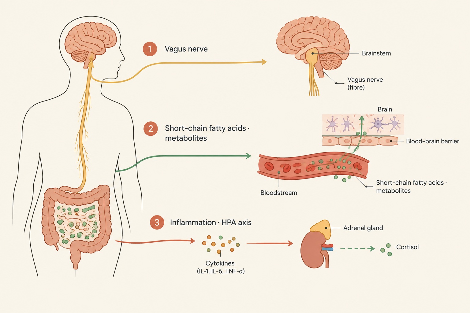
- Figure 2-1 Three routes of the microbiota-gut-brain axis (from the author's lecture notes)
- Figure 2-2 Three routes of the microbiota-gut-brain axis (from the author's lecture notes)
- Figure 2-3 Three routes of the microbiota-gut-brain axis (from the author's lecture notes)
Chapter 1
Introduction
1.1 The problem
The global burden of neuropsychiatric conditions, particularly major depressive disorder and anxiety disorders, presents one of the most pressing challenges to contemporary medicine and public health. For decades, the dominant paradigm in psychiatry has focused on the central monoaminergic hypothesis, which posits that deficits in neurotransmitters such as serotonin, norepinephrine, and dopamine within the synaptic cleft are the primary drivers of depressive and anxious phenotypes. While this framework facilitated the development of first-line pharmacotherapies, including selective serotonin reuptake inhibitors, their clinical efficacy remains suboptimal. A significant proportion of patients exhibit treatment resistance, delayed therapeutic onset, or intolerable side effects, suggesting that the etiology of these disorders extends far beyond localized neurotransmitter imbalances in the brain.
In response to these limitations, psychiatric research has increasingly adopted a systemic, integrative perspective. Depression and anxiety are no longer viewed merely as isolated disorders of the central nervous system, but rather as systemic pathological states characterized by immune dysregulation, endocrine abnormalities, and altered metabolic pathways. Specifically, major depressive disorder is frequently accompanied by a chronic, low-grade inflammatory state, characterized by elevated levels of pro-inflammatory cytokines, cell-mediated immune activation, and heightened oxidative and nitrosative stress (Berk et al., 2013). This inflammatory environment contributes to neuroprogression, a process involving neurodegeneration, decreased neuroplasticity, and altered synaptic connectivity in brain regions regulating mood and cognition (Berk et al., 2013). The identification of these systemic biomarkers has forced a critical re-evaluation of the upstream triggers of chronic inflammation and neuroendocrine dysfunction.
Among the candidate systems capable of modulating both systemic immunity and central nervous system function, the gastrointestinal tract has emerged as a central player. The bidirectional communication network linking the enteric nervous system, the gastrointestinal tract, and the central nervous system—collectively referred to as the gut-brain axis—has long been recognized for its role in maintaining physiological homeostasis (Cryan et al., 2019). However, the discovery that the trillions of microorganisms residing within the human gastrointestinal tract, known as the gut microbiota, actively regulate this axis represents a major paradigm shift in neuroscience and psychiatry (Martin et al., 2018). The microbiota-gut-brain axis integrates neural, hormonal, and immunological signals, allowing microbial communities to influence host behavior, emotional reactivity, and cognitive processes (Cryan et al., 2019).
The fundamental problem confronting contemporary researchers is to elucidate the precise, mechanistic pathways through which the gut microbiome influences the pathophysiology of anxiety and depression. While preclinical models have demonstrated that alterations in the composition and diversity of the gut microbiota can profoundly affect stress reactivity and emotional behavior, translating these findings to human clinical populations remains a complex task. The human gut microbiome is an incredibly diverse and dynamic ecosystem, shaped by genetics, diet, geography, and lifestyle factors, making the identification of specific causal microbial signatures highly challenging. To move beyond simple associations and correlations, researchers must dissect the molecular crosstalk between host and microbe, determine how microbial metabolites cross physiological barriers to reach the brain, and establish how these interactions can be therapeutically targeted to alleviate psychiatric symptoms.
1.2 State of research
Over the past decade, preclinical and clinical investigations have yielded substantial evidence demonstrating the critical role of the gut microbiota in modulating brain function and behavior. Initial insights came from studies utilizing germ-free rodents, which are raised in sterile environments and completely lack a microbiome. These germ-free animals exhibit profound alterations in their neurodevelopment, neurochemistry, and behavior compared to specific-pathogen-free controls. For instance, germ-free rodents display altered anxiety-like behaviors, modified pain perception, and abnormal stress responses, alongside significant alterations in central neurotransmitter systems, particularly in regions such as the amygdala and hippocampus (Mayer et al., 2015). Furthermore, the administration of specific probiotic strains or antibiotic cocktails to adult animals has been shown to dynamically modulate these behavioral and neurochemical profiles, confirming that the influence of the microbiota is not limited to early developmental windows but persists throughout the lifespan (Mayer et al., 2015).
Communication along the microbiota-gut-brain axis occurs via several parallel, highly interconnected pathways. The first of these is the neural pathway, primarily mediated by the vagus nerve. The vagus nerve serves as a direct bidirectional communication highway between the gut and the brain, with approximately eighty percent of its fibers being sensory afferents that transmit information from the visceral organs to the central autonomic network (Bonaz et al., 2018). The vagus nerve is highly sensitive to microbial metabolites and gut hormones, allowing it to act as an interoceptive sensor that monitors the state of the gastrointestinal tract and translates these signals into central neurochemical changes (Bonaz et al., 2018). Additionally, activation of the vagal efferent fibers can trigger the cholinergic anti-inflammatory pathway, which suppresses systemic inflammation and protects against tissue-damaging immune responses (Bonaz et al., 2018).
A second critical communication route is the humoral and metabolic pathway, which involves the systemic circulation of microbial-derived signaling molecules. Among the most widely studied of these metabolites are short-chain fatty acids, primarily acetate, propionate, and butyrate, which are produced in the colon through the bacterial fermentation of dietary fibers (Silva et al., 2020). Short-chain fatty acids act as key signaling molecules that interact with host G-protein coupled receptors, modulating metabolic, endocrine, and immune functions (Silva et al., 2020). These metabolites can cross the blood-brain barrier or act indirectly by stimulating the release of gut hormones and neurotransmitters from enteroendocrine cells, thereby influencing neurochemical pathways and neuroplasticity in the brain. Tryptophan metabolism represents another essential metabolic pathway, as the gut microbiota regulates the availability of tryptophan, the essential precursor for serotonin synthesis (Cryan et al., 2019). By shifting tryptophan metabolism away from the serotonin pathway and toward the kynurenine pathway, microbial dysbiosis can lead to the accumulation of neurotoxic metabolites that contribute to depressive symptoms (Cryan et al., 2019)(Foster et al., 2017).
The immune system represents a third major pathway of communication. The gut microbiota plays an indispensable role in training and regulating the host immune system, maintaining the integrity of the intestinal mucosal barrier, and preventing systemic endotoxemia. Under conditions of dysbiosis, the integrity of the intestinal barrier can become compromised, leading to increased permeability, often colloquially termed a leaky gut. This permeability allows bacterial components, such as lipopolysaccharides, to translocate into the systemic circulation, where they trigger a chronic, low-grade inflammatory response (Berk et al., 2013). This systemic inflammation, characterized by circulating pro-inflammatory cytokines, can affect the central nervous system by altering blood-brain barrier permeability, activating microglia, and disrupting neurotransmitter metabolism, ultimately precipitating depressive and anxious behaviors (Berk et al., 2013).
In clinical settings, researchers have sought to characterize the taxonomic and functional alterations of the gut microbiota in patients diagnosed with major depressive disorder and anxiety disorders. High-throughput sequencing technologies have revealed significant differences in the fecal microbiota composition of depressed patients compared to healthy controls. For example, clinical studies have documented alterations in the relative abundance of major bacterial phyla, such as increases in Bacteroidetes, Proteobacteria, and Actinobacteria, alongside a significant reduction in Firmicutes (Jiang et al., 2015). Interestingly, while some clinical studies report decreased microbial diversity in depressed cohorts, others have observed increased alpha-diversity in patients with active depressive symptoms, highlighting the complex, heterogeneous nature of these disorders and the confounding effects of medication, diet, and lifestyle (Jiang et al., 2015). Despite these discrepancies, the therapeutic potential of targeting the microbiome remains a highly promising avenue of research, with clinical trials exploring the efficacy of prebiotics, probiotics, synbiotics, and dietary modifications in alleviating psychiatric symptoms (Martin et al., 2018).
1.3 Structure of the book
This monograph is structured to provide a comprehensive, graduate-level analysis of the scientific evidence linking the gut microbiome to anxiety and depression. By synthesizing the last decade of preclinical discoveries and clinical trials, the text aims to equip early-career researchers and graduate students with a rigorous understanding of the physiological mechanisms, methodological approaches, and therapeutic implications of the microbiota-gut-brain axis. The subsequent chapters are organized logically to transition from theoretical foundations and methodology to core mechanistic questions, ongoing scientific debates, and future directions.
Chapter 2, Concepts and Theory, establishes the theoretical framework of the monograph. This chapter explores the evolutionary biology of host-microbe symbiosis and details how the gut microbiome acts as a dynamic virtual organ that integrates host physiology. It introduces the core concepts of homeostasis, allostasis, and biological networks, explaining how disruptions in microbial ecology can destabilize the systemic equilibrium of the host, leading to neurobiological and behavioral dysfunction.
Chapter 3, Methods and Sources, provides a critical evaluation of the experimental methodologies and analytical tools utilized in microbiota-gut-brain axis research. It reviews the utility and limitations of various animal models, including germ-free rodents, gnotobiotic systems, and antibiotic-induced dysbiosis models. Additionally, the chapter examines the bioinformatic and sequencing technologies employed to profile microbial communities, such as 16S rRNA amplicon sequencing, shotgun metagenomics, and metabolomic profiling of short-chain fatty acids and other microbial metabolites.
Chapter 4, Historical Context, traces the intellectual history of research into the gut-brain axis. It outlines the evolution of psychosomatic medicine, the discovery of the enteric nervous system, and the eventual integration of microbiological science into classical neurobiology. By examining the key paradigms that have shaped our understanding of stress, immunity, and behavior, this chapter highlights how the discovery of the gut microbiota’s regulatory role has transformed contemporary biological psychiatry.
Chapter 5, Core Question I, addresses the primary mechanistic question: how do microbial signals physically reach and influence the brain? This chapter dissects the relative contributions of the neural, endocrine, and immune pathways. It provides a detailed analysis of vagal afferent signaling, the transport of short-chain fatty acids across the blood-brain barrier, and the cascade of events through which systemic inflammation, secondary to intestinal barrier dysfunction, alters central neurochemistry and microglial activation.
Chapter 6, Core Question II, focuses on the therapeutic translation of these mechanistic insights. It evaluates the clinical and preclinical evidence supporting the use of microbiome-targeted interventions for anxiety and depression. The chapter systematically reviews the efficacy of psychobiotics, synbiotics, dietary interventions, and fecal microbiota transplantation, assessing their capacity to restore microbial homeostasis, reduce systemic inflammation, and alleviate psychiatric symptoms.
Chapter 7, Debates and Challenges, confronts the major controversies, contradictions, and methodological bottlenecks in the field. It addresses the translation gap between rodent models and human clinical populations, the confounding variables that complicate clinical trial design—such as diet, physical activity, and concurrent antidepressant medication—and the challenges of establishing causality in human microbiome studies.
Chapter 8, Conclusion, synthesizes the core findings of the monograph and proposes a unified, integrative model of the microbiota-gut-brain axis in psychiatric disease. It outlines critical knowledge gaps and suggests future research directions, emphasizing the need for longitudinal clinical trials, standardized metabolomic protocols, and personalized medicine approaches that leverage the gut microbiome to optimize mental health outcomes.
Chapter 2
Concepts and Theory
2.1 Key concepts
To construct a rigorous scientific inquiry into how the gut microbiota influences anxiety and depression, it is necessary to establish precise definitions of the core biological and ecological concepts. A frequent point of confusion in early-literature is the distinction between the terms microbiota and microbiome. The gut microbiota refers specifically to the structural community of living microorganisms—including bacteria, archaea, fungi, viruses, and microbial eukaryotes—that reside within the gastrointestinal tract (Cryan et al., 2019). In contrast, the gut microbiome represents a broader ecological definition that encompasses not only the physical microorganisms but also their collective genomes, structural elements, and the complex array of metabolic products they produce within the intestinal microenvironment (Cryan et al., 2019). In the context of neuropsychiatric research, investigators are concerned with both the taxonomic composition of the microbiota and the functional capacity of the microbiome, as the chemical outputs of these organisms are what ultimately interface with host physiology.
The communication network through which these entities interact is designated as the microbiota-gut-brain axis. This axis is a bidirectional, multi-systemic communication network that integrates the central nervous system, the autonomic nervous system, the enteric nervous system, and the gastrointestinal tract with the resident microbial communities (Martin et al., 2018). Rather than operating as an autonomous digestive organ, the gastrointestinal tract is now understood to function as a highly sensitive sensory interface. It monitors luminal contents and transmits this information to the brain, while descending central pathways regulate motility, secretion, local blood flow, mucosal nociception, and intestinal immune tone, thereby reshaping the physical and chemical habitat of the gut microbiota (Mayer et al., 2015). The inclusion of the gut microbiota into this classic axis has redefined modern neurobiology, establishing that peripheral microbes are active participants in this bidirectional signaling cascade (Martin et al., 2018).
In clinical and experimental settings, the state of the gut microbiota is often characterized by its diversity and stability. Dysbiosis refers to a persistent perturbation or imbalance in the composition and functional capacity of the microbial community, typically marked by a loss of beneficial taxa, an overgrowth of potentially pathobiont organisms, and a general reduction in ecological resilience (Jiang et al., 2015). To quantify these microbial states, researchers rely on metrics of diversity. Alpha-diversity measures the richness (the number of different species present) and evenness (the relative abundance of those species) within a single biological sample, often calculated using indices such as the Shannon index or Chao1 index (Jiang et al., 2015). Beta-diversity, conversely, quantifies the difference in microbial composition between different samples or experimental cohorts, allowing researchers to determine whether clinical populations, such as those with major depressive disorder, possess distinct microbial signatures compared to healthy control populations (Jiang et al., 2015).
The chemical language of the microbiota-gut-brain axis consists of several key classes of molecules, foremost among which are short-chain fatty acids. Short-chain fatty acids are the primary metabolic end-products of the anaerobic fermentation of dietary fibers and non-digestible carbohydrates by specific colonic bacteria, with acetate, propionate, and butyrate being the most abundant (Silva et al., 2020). These volatile fatty acids serve as essential energy sources for the host epithelium and function as signaling molecules by binding to specific G-protein coupled receptors expressed on immune cells, enteroendocrine cells, and vagal afferents (Silva et al., 2020). Beyond short-chain fatty acids, the metabolic pathways of the essential amino acid tryptophan are highly critical. Tryptophan is the obligatory precursor for the synthesis of serotonin (5-hydroxytryptamine), a neurotransmitter central to the regulation of mood, anxiety, and sleep (Cryan et al., 2019). The gut microbiota plays a major role in determining the systemic availability of tryptophan by modulating its entry into either the serotonin synthesis pathway or the competing kynurenine pathway, a biochemical branch point that is highly sensitive to inflammatory signaling (Foster et al., 2017).
Finally, the concepts of intestinal permeability and systemic low-grade inflammation are fundamental to the pathophysiology of psychiatric disorders. The intestinal barrier is a complex structure composed of a single layer of epithelial cells sealed by tight junction proteins, covered by a protective mucus layer. When dysbiosis occurs, the integrity of these tight junctions can be compromised, leading to a state of increased intestinal permeability, often referred to as a leaky gut (Berk et al., 2013). This structural failure allows the translocation of bacterial components, such as lipopolysaccharides (endotoxins derived from the outer membrane of Gram-negative bacteria), from the intestinal lumen into the lamina propria and portal circulation (Berk et al., 2013). The presence of these bacterial products in the systemic circulation triggers a chronic, low-grade inflammatory response characterized by the activation of peripheral immune cells and the continuous release of pro-inflammatory cytokines, which can ultimately access the central nervous system and disrupt neurochemical homeostasis (Berk et al., 2013).
2.2 Theoretical traditions
The study of the microbiota-gut-brain axis in relation to anxiety and depression has emerged at the intersection of several rich theoretical traditions in psychiatry, immunology, and evolutionary biology. The primary conceptual shift of the past two decades has been the transition from a strictly neurocentric model of psychiatric illness to a systemic, organismal framework. Throughout the mid-to-late twentieth century, biological psychiatry was dominated by the monoamine hypothesis, which conceptualized depression and anxiety as localized neurochemical imbalances within the synaptic clefts of the brain. The contemporary paradigm of the microbiota-gut-brain axis challenges this isolationist view, proposing instead that the brain is deeply embedded within a web of peripheral physiological systems, with the gut microbiome acting as a virtual endocrine organ that continuously tunes central nervous system function (Martin et al., 2018). This systemic perspective integrates gastroenterology, immunology, and endocrinology into a unified framework for understanding mental health (Mayer et al., 2015).
A foundational theoretical pillar of this field is the inflammatory and oxidative stress hypothesis of depression. Historically, depression was viewed primarily as a psychological or neurochemical disorder, but substantial evidence now supports the theory that depression is accompanied by chronic, low-grade systemic inflammation, activation of cell-mediated immunity, and increased oxidative and nitrosative stress (Berk et al., 2013). The theoretical integration of the microbiome into this framework solves a long-standing question in biological psychiatry: what is the primary source of this chronic, low-grade inflammation in patients who lack an obvious physical infection? The gut microbiome provides a highly plausible answer. Under conditions of chronic psychological stress or poor diet, the gut microbiota undergoes dysbiotic shifts that compromise the intestinal epithelial barrier, leading to the systemic translocation of immunogenic bacterial cell wall components (Foster et al., 2017). This persistent endotoxemia keeps the host immune system in a state of mild, chronic activation, driving the production of inflammatory cytokines that cross the blood-brain barrier and alter neurotransmission, neurogenesis, and synaptic plasticity (Berk et al., 2013).
A second major theoretical tradition is the developmental programming hypothesis. This theory posits that early-life events and environmental exposures during critical windows of development establish lifelong physiological set-points, particularly regarding the sensitivity of the hypothalamic-pituitary-adrenal axis to stress (Foster et al., 2017). The gut microbiota is now recognized as a key environmental factor in this developmental programming. During the perinatal period, the infant gut is colonized by maternal and environmental microbes, a process that coincides with the rapid maturation of the host immune system and the central nervous system (Cryan et al., 2019). Theoretical models based on germ-free rodent studies suggest that without exposure to these early microbial signals, the neural circuits responsible for regulating the stress response do not mature normally, leading to a permanent state of hypothalamic-pituitary-adrenal axis hyper-reactivity and altered emotional behavior in adulthood (Mayer et al., 2015). This developmental framework highlights the existence of sensitive periods during which microbial diversity is required to program healthy neurodevelopmental trajectories (Foster et al., 2017).
Lastly, the interoceptive and autonomic nervous system tradition provides the anatomical and physiological basis for real-time gut-to-brain communication. Interoception is defined as the process by which the nervous system senses, interprets, and integrates signals originating from the internal organs of the body, providing a moment-to-moment map of internal physiological state. Within this tradition, the vagus nerve is conceptualized as the primary interoceptive highway linking the visceral organs to the central autonomic network (Bonaz et al., 2018). The theoretical framework of vagal interoception has been expanded to include the sensing of the gut microbiota and its metabolic state. Vagal afferent fibers, which densely innervate the mucosal and muscular layers of the gut, express a wide array of receptors capable of detecting microbial metabolites, gut hormones, and immune mediators (Bonaz et al., 2018). This allows the brain to receive a continuous stream of sensory input regarding the metabolic and immunological status of the gastrointestinal tract, which is integrated into higher-order brain regions that regulate mood, fear, and affective states (Cryan et al., 2019).
2.3 Analytical framework
To analyze the highly complex, bidirectional pathways through which the gut microbiome influences anxiety and depression, researchers require a structured analytical framework. This framework organizes the vast array of physiological interactions into three main, interconnected pathways: the neural pathway, the humoral and endocrine pathway, and the metabolic and immune pathway. By dissecting the microbiota-gut-brain axis into these discrete yet overlapping channels, we can systematically trace how localized alterations in the colonic lumen translate into alterations in central neurochemistry and emotional behavior.
The neural pathway is primarily mediated by the direct connections between the enteric nervous system and the central nervous system, with the vagus nerve serving as the principal conduit. The vagus nerve is a mixed cranial nerve containing approximately 80% sensory afferent fibers and 20% motor efferent fibers, making it uniquely suited for rapid, bidirectional communication (Bonaz et al., 2018). On the afferent side, vagal fibers in the gut wall do not cross the epithelial barrier to directly contact the luminal contents; instead, they terminate in close proximity to enteroendocrine cells and mucosal immune cells (Cryan et al., 2019). This anatomical positioning allows them to sense microbial metabolites, such as short-chain fatty acids, as well as gut peptides like cholecystokinin and glucagon-like peptide-1, which are released by enteroendocrine cells in response to microbial stimulation (Foster et al., 2017). These chemical signals are converted into electrical impulses that travel up the vagus nerve to the nucleus of the solitary tract in the brainstem, which subsequently projects to the parabrachial nucleus, the thalamus, the amygdala, the hippocampus, and the prefrontal cortex—regions that collectively form the central networks governing fear, anxiety, and emotional processing (Bonaz et al., 2018). On the efferent side, the vagus nerve participates in the cholinergic anti-inflammatory pathway, through which central signals can suppress peripheral inflammation by releasing acetylcholine, which binds to alpha-7 nicotinic acetylcholine receptors on macrophages, inhibiting their release of pro-inflammatory cytokines and thereby protecting the integrity of the intestinal barrier (Bonaz et al., 2018).
The humoral and endocrine pathway operates on a slower, systemic timescale and is centered on the hypothalamic-pituitary-adrenal axis and systemic circulation. When an organism perceives a psychological or physical stressor, the hypothalamus releases corticotropin-releasing hormone, which stimulates the anterior pituitary gland to secrete adrenocorticotropic hormone, ultimately triggering the release of glucocorticoids (such as cortisol in humans or corticosterone in rodents) from the adrenal cortex (Foster et al., 2017). Systemic cortisol exerts profound effects on the gastrointestinal tract, including the alteration of gut motility, the reduction of mucus secretion, and the disruption of epithelial tight junctions, which collectively alter the physical niche available to the microbiota and can induce rapid dysbiosis (Mayer et al., 2015). Conversely, the gut microbiota regulates this endocrine response. Microbial metabolites can influence the synthesis of neurotransmitters and their precursors within the gut (Cryan et al., 2019). Specifically, the microbiota regulates the kynurenine pathway of tryptophan metabolism. Under normal conditions, dietary tryptophan is absorbed and utilized for protein synthesis or converted into serotonin (Cryan et al., 2019). However, under conditions of stress or inflammation, the enzymes tryptophan 2,3-dioxygenase and indoleamine 2,3-dioxygenase are upregulated, diverting tryptophan down the kynurenine pathway (Foster et al., 2017). This diversion not only reduces the systemic availability of tryptophan for central serotonin synthesis but also generates neuroactive kynurenine metabolites, such as quinolinic acid, which can cross the blood-brain barrier and exert neurotoxic effects by activating N-methyl-D-aspartate receptors, contributing to the neurobiological changes observed in major depressive disorder (Cryan et al., 2019).
The metabolic and immune pathway represents the direct interface between microbial biochemistry and host systemic physiology. The primary mediators of this pathway are short-chain fatty acids and bacterial structural components. Short-chain fatty acids produced by microbial fermentation (acetate, propionate, and butyrate) enter the portal vein and systemic circulation, where they exert widespread physiological effects (Silva et al., 2020). Butyrate serves as the primary energy substrate for colonic epithelial cells, promoting their proliferation and the expression of tight junction proteins, which prevents the translocation of immunogenic material (Silva et al., 2020). Furthermore, short-chain fatty acids can cross the blood-brain barrier, where they help maintain its structural integrity and influence the activation state of microglia, the resident immune cells of the brain (Silva et al., 2020). When dysbiosis leads to a reduction in short-chain fatty acid production and a corresponding increase in intestinal permeability, bacterial lipopolysaccharides translocate into the systemic circulation (Berk et al., 2013). This systemic endotoxemia triggers the activation of toll-like receptor 4 on peripheral immune cells, initiating an inflammatory cascade that leads to the systemic release of pro-inflammatory cytokines, including tumor necrosis factor-alpha, interleukin-1 beta, and interleukin-6 (Berk et al., 2013). These peripheral cytokines can signal to the brain through several pathways: they can directly enter the brain through the circumventricular organs, where the blood-brain barrier is naturally deficient; they can bind to cytokine receptors on vagal afferents, sending inflammatory signals to the brainstem; or they can actively alter the transport properties of the blood-brain barrier, leading to localized neuroinflammation, impaired neurogenesis, and the development of depressive and anxious phenotypes (Cryan et al., 2019)(Berk et al., 2013).
By utilizing this multi-pathway analytical framework, researchers can map specific microbial alterations to distinct physiological and behavioral outcomes. For example, a reduction in specific butyrate-producing taxa can be analyzed not merely as a taxonomic shift, but as a multi-step cascade: a decrease in butyrate leads to compromised epithelial tight junctions, which increases lipopolysaccharide translocation, driving systemic inflammation and the activation of the kynurenine pathway, which ultimately reduces central serotonin synthesis and promotes neurotoxicity, manifesting behaviorally as anxiety and depression. This integrated framework provides the necessary mechanistic depth to move beyond simple correlations and toward causal models of microbiota-gut-brain communication.
Chapter 3
Methods and Sources
3.1 Sources
The scientific paradigm investigating how the gut microbiome shapes anxiety and depression relies on an interdisciplinary array of empirical sources. Because the microbiota-gut-brain axis operates at the intersection of microbiology, immunology, neuroendocrinology, and psychiatry, researchers must synthesize data from both preclinical animal experiments and clinical human cohorts (Martin et al., 2018). These diverse sources provide complementary insights, balancing the mechanistic control achievable in laboratory environments with the translational validity required to understand human psychopathology.
Preclinical animal models serve as the foundational source of mechanistic data. Among these, germ-free rodents—animals born and reared in sterile isolators to ensure they completely lack any microbial exposure—represent a vital source (Mayer et al., 2015). By comparing these germ-free animals with specific pathogen-free or conventionally colonized counterparts, researchers obtain baseline data on how the absolute absence of microbes alters neurodevelopment, neurotransmitter systems, and emotional behaviors (Mayer et al., 2015). Beyond germ-free models, preclinical researchers utilize pharmacological and environmental interventions as data sources. These include trials involving broad-spectrum antibiotic cocktails designed to deplete the adult microbiome, as well as targeted probiotic or prebiotic administrations (Cryan et al., 2019). Such models allow for the isolation of specific bacterial strains and their direct physiological effects on the host.
Another critical preclinical source is the human-to-rodent fecal microbiota transplantation. In this experimental design, fecal samples collected from patients diagnosed with major depressive disorder or generalized anxiety disorder are transplanted into microbiota-depleted or germ-free rodents. By observing whether the behavioral and physiological traits of the human donors are recapitulated in the recipient animals, these studies provide a unique source of evidence testing the transmissibility of psychiatric phenotypes via microbial communities.
On the clinical side, the primary sources of data are observational and interventional human cohorts. Observational studies typically compare the fecal microbiota of clinically diagnosed psychiatric patients with matched healthy controls (Jiang et al., 2015). Advanced clinical protocols also incorporate longitudinal profiling, capturing samples from patients during active disease states and comparing them to samples obtained after successful therapeutic responses (Jiang et al., 2015). These clinical cohorts are essential for validating preclinical findings, ensuring that the taxonomic shifts and metabolic perturbations observed in rodents are mirrored in actual human disease states.
In addition to taxonomic data, clinical and preclinical investigations rely on systemic physiological and biochemical biomarkers. Immunological data sources are particularly prominent, given the established link between chronic, low-grade systemic inflammation and the pathogenesis of depressive and anxious symptoms (Berk et al., 2013). Researchers collect peripheral blood samples to measure circulating inflammatory cytokines, immune cell profiles, and markers of cell-mediated immunity (Berk et al., 2013). Other biochemical sources include metabolic profiles of blood, urine, and cerebrospinal fluid, with a specific focus on tryptophan metabolites along the kynurenine pathway and microbial-derived short-chain fatty acids (Silva et al., 2020)(Foster et al., 2017). Finally, neuroimaging databases, such as functional magnetic resonance imaging in humans, and electrophysiological recordings of vagal nerve activity in animals, provide functional readouts of brain-gut connectivity (Bonaz et al., 2018)(Martin et al., 2018).
3.2 Methods
To extract meaningful insights from these diverse sources, researchers employ a sophisticated suite of methodological tools designed to characterize microbial communities, assess behavioral phenotypes, and map bidirectional communication pathways.
3.2.1 Metagenomic and Taxonomic Profiling
The characterization of the gut microbiota relies heavily on high-throughput DNA sequencing. The most widely utilized method is 16S ribosomal RNA gene sequencing, which targets hypervariable regions of the bacterial 16S rRNA gene to identify and classify microbial taxa. Historically, high-throughput pyrosequencing was employed to analyze fecal samples, revealing distinct taxonomic shifts in major phyla—such as increases in Bacteroidetes, Proteobacteria, and Actinobacteria, alongside decreases in Firmicutes—in patients suffering from major depressive disorder (Jiang et al., 2015). In contemporary research, this has been largely augmented by shotgun metagenomic sequencing, which sequences all genomic DNA in a sample. This approach allows for higher-resolution species- and strain-level identification, while also profiling the functional genetic potential of the microbiome.
Bioinformatics pipelines process these sequencing data to calculate ecological diversity metrics. Alpha-diversity, which measures the richness and evenness of species within a single sample, is commonly quantified using indices such as the Shannon index (Jiang et al., 2015). Beta-diversity, which measures the taxonomic variation between different samples or experimental groups, is visualized using principal coordinate analysis based on phylogenetic distance metrics. These computational methods enable researchers to identify specific microbial signatures associated with psychiatric states and correlate them with clinical severity scores.
3.2.2 Preclinical Behavioral Assays
In animal models, evaluating the functional impact of the microbiome on anxiety and depression requires standardized behavioral paradigms. Anxiety-like behavior in rodents is typically assessed using assays that exploit their natural conflict between exploring novel environments and avoiding open, brightly lit spaces. The elevated plus maze and the open field test are the standard methodologies for this purpose. In the elevated plus maze, highly anxious rodents spend significantly less time in the open arms, preferring the safety of the enclosed arms. In the open field test, anxiety is quantified by thigmotaxis, where the animal avoids the center of the arena and remains close to the walls.
Depressive-like behaviors, particularly behavioral despair and anhedonia, are measured using separate paradigms. The forced swim test and the tail suspension test assess behavioral despair, where the duration of immobility is recorded as a proxy for helplessness. Anhedonia, a core symptom of depression, is evaluated using the sucrose preference test, where a reduced preference for a sweetened sucrose solution over plain water indicates a deficit in reward processing. By applying these behavioral assays to germ-free, antibiotic-treated, or probiotic-supplemented rodents, researchers can quantify how specific microbial manipulations alter emotional reactivity and motivational states (Mayer et al., 2015).
3.2.3 Neuroendocrine and Immunological Assays
Mapping the communication pathways between the gut and the brain requires precise neuroendocrine and immunological methods. The reactivity of the hypothalamic-pituitary-adrenal axis, a central regulator of the stress response, is evaluated by measuring circulating levels of corticosterone in rodents or cortisol in humans following exposure to acute stressors (Foster et al., 2017).
Immunological methods are critical for investigating the inflammatory hypothesis of depression (Berk et al., 2013). Researchers utilize enzyme-linked immunosorbent assays and multiplex bead arrays to quantify peripheral and central inflammatory markers, including tumor necrosis factor-alpha, interleukin-6, and interleukin-1 beta. In animal models, immunohistochemistry is employed to visualize microglial activation and morphological changes in brain regions associated with emotional regulation, such as the hippocampus and the amygdala.
Furthermore, the kynurenine pathway of tryptophan metabolism is analyzed using high-performance liquid chromatography or mass spectrometry. Because tryptophan is the essential precursor to serotonin, and its diversion into the kynurenine pathway is driven by inflammatory cytokines, measuring the ratio of kynurenine to tryptophan in plasma or brain tissue reveals whether gut-derived inflammation is compromising central serotonin synthesis (Cryan et al., 2019).
3.2.4 Physiological and Neurological Tracking
Identifying the physical routes of communication between the gut and the central nervous system requires targeted physiological interventions. The vagus nerve is a major bidirectional pathway in the microbiota-gut-brain axis (Bonaz et al., 2018). To study its involvement, researchers perform surgical subdiaphragmatic vagotomies in rodents prior to microbial interventions. If the behavioral or neurochemical effects of a probiotic are abolished following this surgery, it demonstrates that the bacterial signal is transmitted to the brain via vagal afferents (Cryan et al., 2019).
Electrophysiological methods are also used to record afferent vagal nerve activity in real-time. By placing electrodes on the vagus nerve, researchers can measure changes in action potential firing rates in response to the luminal perfusion of specific bacterial metabolites, such as short-chain fatty acids or gut peptides (Bonaz et al., 2018). Additionally, pharmacogenetic tools, such as designer receptors exclusively activated by designer drugs, are increasingly used to selectively activate or silence specific enteric neurons or vagal afferents, providing precise mechanistic validation.
3.2.5 Metabolomic Profiling
To decode the chemical signaling of the microbiome, researchers utilize metabolomic profiling. This is highly relevant for studying short-chain fatty acids, which are the primary metabolites produced by the bacterial fermentation of dietary fibers in the colon (Silva et al., 2020). Gas chromatography-mass spectrometry and liquid chromatography-mass spectrometry are employed to detect and quantify short-chain fatty acids, such as acetate, propionate, and butyrate, in fecal, cecal, portal, and systemic blood samples (Silva et al., 2020). These methods allow researchers to correlate specific microbial metabolic outputs with physiological changes in the host, such as blood-brain barrier integrity, microglia maturation, and the release of gut hormones (Silva et al., 2020).
3.3 Limitations
While the integration of these methods has significantly advanced the understanding of the microbiota-gut-brain axis, several methodological, translational, and conceptual limitations must be considered.
3.3.1 Preclinical to Clinical Translational Barriers
The heavy reliance on rodent models introduces substantial translational challenges. Although rodents share many physiological similarities with humans, their gastrointestinal anatomy, metabolic rates, and baseline microbial compositions differ significantly. For example, rodents possess a large cecum where the bulk of bacterial fermentation occurs, whereas in humans, fermentation is distributed throughout the colon. Furthermore, the rodent gut microbiota is distinct from the human microbiota at the genus and species levels, meaning that findings regarding specific bacterial strains in mice may not directly apply to human biology.
The behavioral assays used to model anxiety and depression in rodents also have inherent limitations. Despair-based tests like the forced swim test have been criticized for measuring adaptive energy-conserving behavior rather than true depressive despair. Human depression and anxiety are highly complex, heterogeneous psychiatric conditions characterized by subjective emotional states, cognitive distortions, and verbal communication, none of which can be fully captured by immobility in water or avoidance of open spaces (Mayer et al., 2015).
Furthermore, germ-free rodents, while invaluable for establishing causality, represent an extreme and highly artificial physiological state. These animals exhibit profound developmental abnormalities, including an immature immune system, altered brain development, compromised blood-brain barrier integrity, and exaggerated hypothalamic-pituitary-adrenal axis reactivity (Mayer et al., 2015)(Foster et al., 2017). Consequently, observing a phenotype in a germ-free mouse may reflect a developmental defect resulting from the lifelong absence of microbes, rather than a direct, acute signaling mechanism that can be targeted in adult humans.
3.3.2 Clinical Confounders and Heterogeneity
Clinical studies of the gut microbiota in psychiatric populations are frequently complicated by high inter-individual variability and numerous confounding factors. One of the most significant confounders is diet. Diet is a primary driver of gut microbiota composition, capable of rapidly altering microbial populations and their metabolic outputs (Foster et al., 2017). In free-living human cohorts, accurately tracking and controlling dietary intake is notoriously difficult. Differences in fiber consumption, macronutrient ratios, and the intake of fermented foods or artificial sweeteners can easily obscure or mimic disease-specific microbial signatures.
Medication use represents another formidable confounder. Many patients with major depressive disorder or anxiety disorders are taking psychotropic medications, such as selective serotonin reuptake inhibitors, which have been shown to possess antimicrobial properties and directly alter the composition of the gut microbiota (Jiang et al., 2015). Conversely, other common medications, including proton pump inhibitors, laxatives, and antibiotics, exert profound effects on the microbiome. Distinguishing between dysbiosis associated with the underlying psychiatric pathology and dysbiosis induced by pharmacological treatment remains a major hurdle, even when comparing active and responded patient groups (Jiang et al., 2015).
Other lifestyle and demographic variables—such as age, sex, body mass index, sleep patterns, physical activity, and geographic location—also shape the microbiome and are rarely fully matched or controlled for in clinical trials. Additionally, depression is highly comorbid with systemic inflammatory conditions, metabolic syndrome, and gastrointestinal disorders like irritable bowel syndrome. Because systemic inflammation itself can alter both brain function and gut barrier permeability, determining the primary driver of dysbiosis in these complex, multi-systemic disorders is exceedingly difficult (Berk et al., 2013).
3.3.3 Methodological and Technological Constraints
The methods used to analyze the microbiome also introduce significant biases. Fecal samples are the standard source of microbial DNA in human studies due to their non-invasive collection. However, fecal microbiota is primarily representative of the luminal environment of the distal colon. It does not accurately reflect the microbial communities of the small intestine, nor does it capture the mucosal-associated microbiota that directly interacts with the host's intestinal epithelium and mucosal immune system.
Furthermore, the reliance on 16S rRNA gene sequencing limits the depth of taxonomic resolution. This method often cannot resolve bacteria down to the species or strain level, which is critical because different strains within the same species can have vastly different functional properties and physiological impacts on the host. While shotgun metagenomics addresses this by sequencing all genomic DNA, it is significantly more expensive and computationally demanding.
Additionally, ecological metrics like alpha-diversity can yield contradictory findings. For instance, while some clinical studies report decreased microbial diversity in depressed patients, others have observed increased alpha-diversity in active-depressive patients compared to healthy controls (Jiang et al., 2015). These discrepancies may stem from differences in sequencing depth, bioinformatics pipelines, patient demographics, or sample handling protocols, highlighting the lack of standardization across the field.
3.3.4 The Correlation-Causation Dilemma
Perhaps the most fundamental limitation of current clinical evidence is its cross-sectional nature. Most human studies profile the microbiome at a single time point, demonstrating associations between specific microbial taxa and psychiatric symptoms but failing to establish directionality (Martin et al., 2018). It remains unclear whether alterations in the gut microbiota contribute to the pathogenesis of anxiety and depression, or whether the physiological state of being depressed or anxious—characterized by high hypothalamic-pituitary-adrenal axis activity, altered autonomic nervous system output, and poor dietary choices—subsequently alters the gut environment and shapes the microbiota (Foster et al., 2017). Longitudinal studies, interventional trials, and mechanistic animal models are continuously required to untangle this bidirectional relationship and move the field from correlation to true causal understanding (Cryan et al., 2019)(Martin et al., 2018).
Chapter 4
Historical Context
4.1 Early work
The proposition that emotional states are tethered to the viscera is among the oldest observations in medicine, yet for over a century it remained marooned between poetic clinical intuition and ungrounded speculation. In the late nineteenth and early twentieth centuries, alienists and physicians frequently observed a relationship between gastrointestinal disturbance and melancholic syndromes. This intuition took clinical shape in the theory of autointoxication, which posited that stagnant fecal material in the colon generated putrefactive poisons that circulated to the brain, producing mental confusion, fatigue, and profound sadness. Without rigorous microbiological techniques or an understanding of mucosal immunology, however, the autointoxication doctrine degenerated into therapeutic excess. Patients were subjected to aggressive colonic irrigations, restrictive regimens, and even surgical colectomies to rid the body of presumptive internal poisons. When controlled pathology failed to corroborate these claims, the medical establishment rightfully discarded the premise. The subsequent rise of psychoanalysis on the one hand and the monoaminergic revolution in psychopharmacology on the other relegated the digestive tract to an incidental status, viewing visceral distress merely as a peripheral byproduct of central psychiatric disturbance.
Throughout the middle decades of the twentieth century, gastrointestinal physiology and neurobiology progressed along largely separate tracks. Enteric neuroscience established that the bowel possessed its own vast, semi-autonomous nervous system capable of orchestrating peristalsis, fluid transport, and local blood flow without continuous central instruction. Meanwhile, autonomic physiology mapped the pathways of bidirectional communication connecting the brainstem to the gut, demonstrating how emotional stress altered motility and mucosal secretions. Yet in these formulations, the luminal contents were treated as an inert chemical slurry or a collection of opportunistic pathogens held at bay by the intestinal epithelium. The organisms inhabiting the intestine were recognized for their capacity to synthesize vitamin K and break down otherwise indigestible plant polysaccharides, but they were not considered active regulators of neural development or affective behavior. Gastrointestinal medicine focused on eradication of overt pathogens, while biological psychiatry focused almost exclusively on monoamine neurotransmission, synaptic receptor density, and hypothalamic control. The resident community of microbes remained physiologically invisible to the neurosciences (Cryan et al., 2019)(Mayer et al., 2015).
The experimental tools that would eventually bridge this divide originated not in psychiatry, but in the field of gnotobiology. During the mid-twentieth century, researchers developed sterile isolator technology that made it possible to derive and rear germ-free mice across multiple generations. Initially, GF mice were deployed to answer fundamental questions in nutrition, immunology, and infectious disease, such as whether vertebrate life could be sustained in the absolute absence of microbial colonization and how the host immune system developed its primary architecture. These early investigations revealed substantial anatomical and physiological aberrations in germ-free mice: an enlarged cecum, altered mucosal vascularization, defective development of gut-associated lymphoid tissue, and distinct metabolic shifts. Nevertheless, for decades behavioral scientists paid little attention to these animals. The central nervous system was assumed to be shielded from the luminal environment by both the intestinal epithelial barrier and the blood-brain barrier, leaving little conceptual space for intestinal microbes to exert any meaningful influence over complex brain function.
A parallel development in somatic medicine gradually created the empirical conditions necessary to re-evaluate peripheral inputs to the brain: the study of sickness behavior and systemic inflammation. By the late 1980s and 1990s, neuroimmunologists demonstrated that the systemic administration of lipopolysaccharide or pro-inflammatory cytokines such as interleukin-1 and tumor necrosis factor elicited a stereotypic constellation of behavioral changes in rodents, including lethargy, anhedonia, decreased social exploration, and anorexia. Far from being a passive manifestation of toxic debilitation, this state represented an organized central motivational shift mediated by cytokine signaling across the blood-brain barrier and along neural routes. This realization prompted psychiatric researchers to formulate the inflammatory hypothesis of depression, observing that a significant subset of patients with major depressive disorder exhibited persistent elevations in circulating inflammatory markers, oxidative stress, and cell-mediated immune activation (Berk et al., 2013). Yet this hypothesis immediately faced an unresolved mechanistic dilemma: in the absence of an active autoimmune disorder or an overt systemic infection, the primary tissue source of this chronic low-grade inflammation remained unidentified (Berk et al., 2013).
4.2 Turning points
The decisive shift from viewing intestinal microbes as passive commensals to recognizing them as active regulators of neurobiology occurred at the turn of the twenty-first century. The foundational turning point was established in 2004, when Sudo and colleagues examined the neuroendocrine stress response in germ-free mice (Mayer et al., 2015)(Foster et al., 2017). When subjected to mild restraint stress, adult GF mice exhibited an exaggerated elevation of plasma adrenocorticotropic hormone and corticosterone compared to specific-pathogen-free mice reared with a standard gut microbiota (Mayer et al., 2015)(Foster et al., 2017). Crucially, this hyper-reactive stress response was partially reversed by reconstituting the intestine with a single commensal organism, Bifidobacterium infantis, or fully normalized by colonization with fecal suspensions from control animals, but only if the colonization took place at an early developmental stage (Mayer et al., 2015)(Foster et al., 2017). Reconstituting adult germ-free mice failed to correct the abnormal endocrine profile, revealing for the first time that signals originating from the gut microbiota are indispensable for the programmed developmental maturation of the hypothalamic-pituitary-adrenal axis (Mayer et al., 2015)(Foster et al., 2017).
Following the demonstration of neuroendocrine vulnerability, research groups in the early 2010s set out to establish whether the absence or alteration of the gut microbiota also affected emotional and exploratory behavior. Independent laboratories confirmed that germ-free mice displayed altered anxiety-like behavioral phenotypes in standard paradigms such as the elevated plus maze, the light-dark box, and the open field test (Mayer et al., 2015)(Foster et al., 2017). In several mouse strains, GF mice exhibited reduced anxiety-like behavior and increased motor exploration, which coincided with alterations in central neurochemistry, including modified expression of brain-derived neurotrophic factor in the hippocampus and amygdala, along with widespread shifts in monoamine turnover rates (Mayer et al., 2015)(Foster et al., 2017). While the directionality of behavioral phenotypes varied depending on host genetics and testing protocols, the underlying principle was established: host neurodevelopment and baseline behavioral traits are sensitive to the presence of luminal microorganisms during critical windows of ontogeny (Mayer et al., 2015).
Once behavioral phenotypes were documented in germ-free mice, investigators turned to the anatomical and biochemical routes mediating this dialogue within the microbiota-gut-brain axis (Cryan et al., 2019). Attention focused rapidly on the vagus nerve, which serves as the principal direct physical link between the viscera and the central nervous system. Anatomical tracing confirmed that approximately eighty percent of vagal fibers are afferent, terminating in the nucleus of the solitary tract in the brainstem, from which signals ascend to the central autonomic network and limbic structures regulating emotional processing (Bonaz et al., 2018). Experimental work demonstrated that the ingestion of specific probiotic bacterial strains produced changes in central gamma-aminobutyric acid receptor expression and reduced anxiety- and depressive-like behaviors in rodents, effects that were completely abolished when the subdiaphragmatic vagus nerve was surgically severed (Bonaz et al., 2018). The efferent vagal fibers were concurrently identified as mediators of the cholinergic anti-inflammatory pathway, which dampens the systemic release of pro-inflammatory cytokines and reinforces mucosal barrier integrity, establishing the vagus nerve as a bidirectional highway in the microbiota-gut-brain axis (Bonaz et al., 2018).
Simultaneously, metabolic profiling identified short-chain fatty acids as critical humoral messengers linking microbial fermentation to host neurophysiology (Silva et al., 2020). Acetate, propionate, and butyrate, produced primarily by anaerobic taxa in the colon through the fermentation of complex non-digestible carbohydrates, were shown to transcend their classical roles as energy substrates for colonocytes (Silva et al., 2020). Pharmacological and physiological investigations revealed that these short-chain fatty acids activate specific cell-surface G-protein-coupled receptors and function as endogenous inhibitors of histone deacetylases (Silva et al., 2020). Through these mechanisms, circulating SCFAs help preserve intestinal barrier integrity, cross the blood-brain barrier via monocarboxylate transporters, and regulate the maturation and transcriptional profile of microglia, the resident immune cells of the central nervous system (Silva et al., 2020). This metabolic discovery provided a concrete biochemical mechanism by which dietary changes and shifts in bacterial fermentation could alter central neuroinflammatory tone and behavior (Silva et al., 2020).
The conceptual leap from correlation to functional transmissibility came with the adoption of fecal microbiota transplantation in behavioral animal models (Cryan et al., 2019)(Martin et al., 2018). Researchers demonstrated that behavioral traits could be transferred between distinct rodent strains solely by exchanging their gut microbiota through gavage into recipient animals. More provocatively, subsequent preclinical studies showed that colonizing antibiotic-depleted or germ-free rodents with fecal material obtained from human donors diagnosed with major depressive disorder induced depressive- and anxiety-like behaviors in the recipient animals, characterized by anhedonia in the sucrose preference test and increased immobility in the forced swim test (Martin et al., 2018). These recipient animals also recapitulated several metabolic and immune perturbations seen in the human disorder, such as altered tryptophan metabolism and heightened peripheral inflammation (Cryan et al., 2019)(Martin et al., 2018). While questions remained regarding the degree to which complex human mood symptoms can be accurately modeled in rodents, these fecal microbiota transplantation experiments established a vital proof-of-concept: microbial consortia can act as causal mediators of behavioral phenotypes rather than just passive markers of disease (Martin et al., 2018).
4.3 The present
Over the past decade, high-throughput DNA sequencing transformed the study of the microbiota-gut-brain axis from a specialized preclinical niche into a prominent frontier of human psychiatric neuroscience (Cryan et al., 2019)(Martin et al., 2018). The rapid drop in sequencing costs allowed researchers to abandon culture-dependent assays in favor of 16S rRNA gene sequencing and shotgun metagenomics, making large-scale clinical surveys of psychiatric cohorts feasible (Jiang et al., 2015). Cross-sectional investigations profiling stool samples from individuals with major depressive disorder consistently identified compositional differences compared to healthy controls (Jiang et al., 2015). Clinical studies reported distinct shifts in taxonomic abundance, including reductions in taxa producing short-chain fatty acids and alterations in overall community diversity, with several studies documenting that the magnitude of these compositional deviations correlated with the severity of depressive and anxiety symptoms (Jiang et al., 2015). These human datasets corroborated the preclinical consensus that psychiatric morbidity is accompanied by disrupted microbial ecology in the intestinal lumen (Jiang et al., 2015)(Martin et al., 2018).
Modern models of the microbiota-gut-brain axis integrate these findings into an interconnected tripartite architecture comprising neural, metabolic, and neuroimmune routes (Cryan et al., 2019). The neuroimmune route has provided a direct answer to the longstanding question of where systemic inflammation in major depressive disorder originates (Berk et al., 2013). Under homeostatic conditions, the gut microbiota maintains intestinal epithelial tight junctions and educates host immune responses. When dysbiosis occurs, elevated intestinal permeability allows bacterial cell-wall fragments, most notably lipopolysaccharide, to translocate into the lamina propria and the systemic circulation (Berk et al., 2013). This endotoxemia engages toll-like receptors on circulating immune cells, precipitating a low-grade, chronic systemic inflammatory response characterized by elevated circulating levels of interleukin-1beta, interleukin-6, and tumor necrosis factor-alpha (Berk et al., 2013). These pro-inflammatory cytokines compromise the blood-brain barrier, promote the activation of microglia, and stimulate the enzyme indoleamine 2,3-dioxygenase, which diverts systemic and central tryptophan metabolism away from serotonin synthesis and toward the neurotoxic branches of the kynurenine pathway (Berk et al., 2013).
``` +-----------------------------+ | Host Stresses and | | Environmental Factors | +--------------+--------------+ | v +-----------------------------+ | Intestinal Dysbiosis | +--------------+--------------+ | +-------------------------+-------------------------+ | | v v +-----------------------+ +-----------------------+ | Barrier Breakdown: | | Metabolic & Neural | | LPS Translocation | | Pathways: | +----------+------------+ | Altered SCFA levels | | | and Vagal signaling | v +-----------+-----------+ +-----------------------+ | | Systemic Inflammation | | | (IL-1b, IL-6, TNF-a) | | +----------+------------+ | | | +-------------------------+-------------------------+ | v +-----------------------------+ | Microglia Activation | | and Altered Neuroplasticity| +--------------+--------------+ | v +-----------------------------+ | Altered Affect and | | Mood-Related Behaviors | +-----------------------------+ ```
Despite the elegance of these mechanistic pathways, the contemporary literature faces significant methodological frictions and interpretive challenges. The overwhelming majority of human studies linking gut microbiota composition to major depressive disorder and anxiety disorders remain cross-sectional in design, capturing a single taxonomic snapshot in time (Jiang et al., 2015). Stool composition is exceptionally labile, responding acutely to diet, physical exercise, tobacco use, systemic stress, and sleep deprivation, as well as to pharmacological agents. Antidepressants, sedatives, antipsychotics, and non-psychiatric medications such as proton pump inhibitors exert potent antimicrobial effects, confounding observed associations between microbial taxa and clinical diagnostic categories. High-throughput 16S rRNA gene sequencing yields compositional proportions of bacterial operational taxonomic units, which do not necessarily reflect absolute microbial loads or functional metabolic output (Jiang et al., 2015). Consequently, an observed taxon shift in a depressed cohort may represent a downstream consequence of altered appetite, medication use, or lifestyle rather than an upstream driver of affective pathology.
The translational frontier is currently defined by interventional trials attempting to alter emotional states through targeted manipulation of the gut microbiota (Cryan et al., 2019)(Martin et al., 2018). Preclinical models established that specific living microorganisms, broadly categorized as psychobiotics, can downregulate HPA axis reactivity and reduce anxiety-related exploratory deficits (Cryan et al., 2019)(Foster et al., 2017). Clinical translation, however, has produced a heterogeneous evidentiary record. Randomized controlled trials evaluating multi-strain probiotic combinations, prebiotic dietary fibers, and whole-diet modifications in populations with major depressive disorder and generalized anxiety have reported modest or mixed outcomes. While certain trials report significant reductions in self-reported depressive scores and inflammatory biomarkers, others find no distinction from placebo. The emerging field of therapeutic fecal microbiota transplantation for treatment-resistant affective disorders remains similarly experimental, governed by small sample sizes and variable donor-matching protocols (Martin et al., 2018).
The history of the microbiota-gut-brain axis has thus evolved through three major epochs: from unverified somatic autointoxication, through mechanistic proof-of-concept in controlled animal models, to the present era of high-throughput clinical association and interventional trials (Cryan et al., 2019)(Martin et al., 2018). The fundamental challenge facing current investigators is epistemological discipline. The field has moved past the need to prove that communication exists between the gut microbiota and the central nervous system; the existence of the neural, endocrine, and immune routes of the MGBA is now an established physiological reality (Cryan et al., 2019). The task ahead is to replace broad descriptive correlations with rigorous, prospective, and mechanistically defined clinical studies that differentiate true microbial causation from incidental collateral shifts, clarifying precisely how, and to what extent, the gut microbiota shapes human anxiety and depression.
Chapter 5
Core Question I
5.1 Question and evidence
The fundamental question of this chapter concerns directionality: does the gut microbiota operate as a primary causal driver of emotional dysregulation, anxiety, and depressive symptoms, or do microbial shifts simply reflect the downstream physiological consequences of affective illness? In the popular imagination and much of the early literature, the gut is often cast as an upstream conductor of central neural processes, suggesting that perturbations within the enteric ecosystem are sufficient to induce psychiatric phenotypes. Yet in human clinical research, emotional disturbance coincides with systemic changes in host behavior, neuroendocrine output, autonomic balance, and diet. Determining whether the gut microbiota acts as an initiator of mood pathology or as a passenger of host distress requires separating correlation from causal mechanism across both animal models and human patient cohorts.
Preclinical exploration of this question began largely with germ-free mice (GF mice), which provide an absolute baseline wherein host biology develops in the total absence of microbial colonization. Pioneering investigations established that GF mice exhibit marked neurodevelopmental and endocrine abnormalities, most prominently an exaggerated reactivity of the hypothalamic-pituitary-adrenal axis (HPA axis) when exposed to acute restraint stress (Mayer et al., 2015)(Foster et al., 2017). When subjected to mild homeostatic challenge, GF mice release substantially higher plasma levels of adrenocorticotropic hormone and corticosterone than specific-pathogen-free controls. Crucially, this hyper-responsive stress profile can be reversed by monocolonization with specific bacterial strains or by reconstitution with a complete gut microbiota, but only if that reconstitution occurs during an early critical developmental window (Foster et al., 2017). Adult colonization leaves the hyper-reactive neuroendocrine phenotype intact, demonstrating that early exposure to microbial signals is necessary for the normative calibration of the central stress response.
Beyond neuroendocrine reactivity, behavioural testing of GF mice revealed consistent alterations in exploratory patterns, locomotion, and anxiety-like traits. In assays such as the elevated plus maze, the light-dark box, and the open field, GF mice typically spend more time in open or brightly illuminated zones, an outcome initially described as anxiolytic-like behaviour, though it may equally reflect cognitive deficits or impaired risk assessment (Cryan et al., 2019)(Mayer et al., 2015). Parallel changes appear in central neurochemistry: GF mice display altered concentrations of brain-derived neurotrophic factor in the hippocampus and amygdala, along with substantial alterations in serotonin turnover and receptor subunit expression throughout the cortex (Cryan et al., 2019). Antibiotic depletion protocols in adult specific-pathogen-free rodents further demonstrate that transient, profound disruption of the gut microbiota can acutely perturb exploratory behaviour and cognitive flexibility, indicating that ongoing microbial activity continues to modulate central processes long after developmental windows have closed.
When investigators moved from sterile models to clinical populations, the primary tool became high-throughput 16S rRNA gene sequencing of fecal samples gathered from psychiatric cohorts. Studies evaluating patients diagnosed with major depressive disorder (MDD) consistently identify significant divergence in microbial composition relative to matched healthy controls (Jiang et al., 2015). Patients experiencing active depressive episodes frequently present with altered bacterial alpha-diversity and distinct shifts in taxonomic abundance, including an elevation of the phyla Bacteroidetes, Proteobacteria, and Actinobacteria, paired with a compensatory contraction of Firmicutes (Jiang et al., 2015). Furthermore, specific family- and genus-level abundances track symptom severity as measured by standardized clinical rating scales (Cryan et al., 2019)(Jiang et al., 2015). The presence of these taxonomic shifts confirms that clinical mood pathology coincides with an altered luminal environment, establishing dysbiosis as a verifiable feature of the depressed state.
To test whether this clinical dysbiosis retains causal efficacy, researchers turned to inter-species fecal microbiota transplantation (FMT). In these experiments, fecal slurries obtained from human donors with major depressive disorder are administered via oral gavage to microbiota-depleted or GF mice (Cryan et al., 2019)(Martin et al., 2018). Across multiple independent laboratories, recipient rodents colonised with stool from depressed individuals exhibited hallmarks of anhedonia, increased immobility during the forced swim test, heightened anxiety-like behaviour in exploratory mazes, and alterations in peripheral lipid and carbohydrate metabolism compared to counterparts colonised with stool from healthy human donors (Martin et al., 2018). These behavioural shifts coincided with changes in central neurotransmitter levels and heightened inflammatory markers, providing empirical support for the hypothesis that the gut microbiota from patients with major depressive disorder carries transmissible pathological properties capable of modifying emotional behaviour across species barriers.
Parallel to fecal transfer paradigms, interventional studies have attempted to alleviate anxiety and depressive symptoms in humans using targeted microbial supplements. Controlled trials of probiotics and prebiotics in subclinical and clinical cohorts demonstrate modest reductions in self-reported psychological distress, reduced cognitive reactivity to sad mood, and attenuated neuroendocrine responses to social stress tasks (Mayer et al., 2015)(Martin et al., 2018). Neuroimaging studies in healthy human volunteers show that chronic ingestion of fermented milk products containing selected bacterial strains alters the reactivity of the central autonomic network and affective limbic circuits during emotional face-matching challenges (Mayer et al., 2015). While these human interventional trials show modest effect sizes and suffer from considerable heterogeneity in bacterial strains and dosing regimens, they corroborate the premise that gut microbiota manipulations can influence emotional processing circuits in the human brain.
The accumulating evidence thus documents a coherent series of observations: total absence of microbes distorts neurodevelopment and stress responsiveness; adult microbial depletion alters affective behaviour; patients with major depressive disorder exhibit measurable dysbiosis; human-to-rodent fecal microbiota transplantation transfers specific behavioural traits; and targeted microbial interventions exert modest regulatory actions on human emotional processing. What remains unresolved is the biological route through which these effects travel, and whether these mechanisms operate with sufficient strength in naturalistic, non-experimental settings to generate depressive and anxiety disorders de novo.
5.2 Analysis
To assess the causal claims embedded in the evidence, one must delineate the physical routes that constitute the bidirectional architecture of the microbiota-gut-brain axis (MGBA). Communication between the enteric lumen and the central nervous system does not occur through ethereal diffusion; it relies upon three well-defined physiological pathways: neural transmission via the autonomic nervous system, metabolic and neuroendocrine signaling driven by microbial fermentation products, and systemic immune-inflammatory cascades (Cryan et al., 2019)(Martin et al., 2018). Each of these routes provides a plausible physical substrate for gut-to-brain signalling, yet each carries distinct mechanistic constraints that limit how far its explanatory power can be extended.
The direct neural route is dominated by the vagus nerve, which serves as the principal neuroanatomical conduit linking the enteric nervous system to the central neuraxis (Cryan et al., 2019)(Bonaz et al., 2018). Structurally, approximately eighty percent of vagus nerve fibres are afferent, originating in the subepithelial layers of the gut wall and projecting directly to the nucleus of the solitary tract in the brainstem (Bonaz et al., 2018). From this primary relay station, ascending projections distribute visceral sensory signals to the locus coeruleus, the parabrachial nucleus, the thalamus, the amygdala, and the insular cortex, thereby interfacing directly with the central autonomic network and the limbic regions that govern emotional valence and anxiety (Bonaz et al., 2018). The remaining twenty percent of vagus nerve fibres are efferent, coordinating motor tone, gastrointestinal motility, glandular secretion, and the cholinergic anti-inflammatory pathway, which dampens peripheral macrophage activation and cytokine release (Bonaz et al., 2018).
Preclinical experiments demonstrate that the vagus nerve is an indispensable link for specific microbial signals. When rodents are subjected to subdiaphragmatic vagotomy, the anxiolytic and behavioural effects of orally administered probiotic strains disappear completely, while sham-operated animals retain the full spectrum of neurochemical and behavioural improvements (Cryan et al., 2019). The physical structure of vagal afferents, however, means they do not penetrate the gut epithelial monolayer into the open lumen. Consequently, the vagus nerve does not sense the gut microbiota directly through physical contact; rather, it responds to microbial metabolites, luminal antigens, and regulatory peptides secreted by enteroendocrine cells, such as cholecystokinin and serotonin, which diffuse into the lamina propria (Bonaz et al., 2018). While vagal afferents possess receptors capable of detecting these chemical cues, vagotomy studies in rodents employ severe, total surgical denervation of subdiaphragmatic branches. Such experiments prove that the intact vagus nerve is necessary for certain probiotic actions in an animal, but they do not prove that natural variations in gut microbiota composition dynamically drive clinical depression in an intact human patient.
The second primary route involves microbial metabolites that enter systemic circulation, most notably short-chain fatty acids (SCFAs). The principal short-chain fatty acids—acetate, propionate, and butyrate—are produced in high concentrations in the colon through the anaerobic fermentation of non-digestible dietary fibre (Silva et al., 2020). At the local level, short-chain fatty acids act as the essential energy substrate for colonocytes, preserve the integrity of intestinal epithelial tight junctions, and bind to cell-surface G-protein-coupled receptors, including free fatty acid receptors 2 and 3 (Cryan et al., 2019)(Silva et al., 2020). A fraction of locally generated short-chain fatty acids escapes splanchnic extraction, enters the systemic bloodstream, and crosses the blood-brain barrier (BBB) via monocarboxylate transporters (Silva et al., 2020).
Once across the blood-brain barrier, short-chain fatty acids exert profound epigenetic and cellular effects within the central nervous system. Butyrate and propionate act as endogenous inhibitors of histone deacetylases (HDACs), modifying gene expression, enhancing the transcription of neurotrophic factors such as brain-derived neurotrophic factor, and supporting neuroplasticity (Silva et al., 2020). Furthermore, short-chain fatty acids are essential for the structural and functional maturation of microglia, the resident macrophages of the central nervous system (Cryan et al., 2019)(Silva et al., 2020). In GF mice, microglia display an immature, malformed morphology and defective transcriptional profiles; this pathology can be rescued by the systemic administration of a physiological mixture of short-chain fatty acids (Cryan et al., 2019). However, physiological concentrations of short-chain fatty acids in human peripheral arterial blood are orders of magnitude lower than those measured in the colonic lumen. Whether circulating concentrations in adult humans fluctuate within a range capable of directly altering central histone acetylation or microglial function remains an open empirical question rather than a settled fact.
The metabolic route also encompasses the regulation of essential amino acid availability, most visibly within tryptophan metabolism. Tryptophan is an indispensable dietary amino acid that serves as the precursor for the synthesis of central serotonin (Cryan et al., 2019)(Foster et al., 2017). The gut microbiota modulates systemic tryptophan availability both by direct utilization and by regulating the enzymatic pathways responsible for its catabolism (Cryan et al., 2019). Under baseline conditions, the majority of dietary tryptophan is metabolized along the kynurenine pathway, initiated by the rate-limiting enzymes tryptophan 2,3-dioxygenase in the liver and indoleamine 2,3-dioxygenase in extrahepatic and immune tissues (Cryan et al., 2019)(Berk et al., 2013). Microbial dysbiosis and associated inflammatory signalling upregulate indoleamine 2,3-dioxygenase, diverting tryptophan away from serotonin production and toward the generation of kynurenine, quinolinic acid, and kynurenic acid (Cryan et al., 2019)(Berk et al., 2013). Quinolinic acid acts as an N-methyl-D-aspartate receptor agonist capable of inducing excitotoxicity and oxidative stress, whereas kynurenic acid acts as an antagonist; shifts in the ratio of these metabolites correlate directly with depressive severity and anhedonia in clinical cohorts (Berk et al., 2013).
The third communication pathway operates through the host immune system and peripheral inflammatory cascades. A stable gut microbiota trains host immune cells, maintains local tolerance, and preserves the mucosal barrier. When dysbiosis occurs, mucosal homeostasis breaks down, tight-junction proteins such as claudin and occludin degrade, and intestinal permeability rises, leading to what is commonly termed a leaky gut (Berk et al., 2013). This structural impairment permits the pathological translocation of luminal bacterial fragments, most notably lipopolysaccharide (LPS), an outer membrane component of Gram-negative bacteria, across the lamina propria and into the portal circulation (Berk et al., 2013). Translocated lipopolysaccharide binds to Toll-like receptor 4 complexes on circulating monocytes and tissue macrophages, triggering an inflammatory cascade characterized by the sustained release of pro-inflammatory cytokines, including interleukin-1 beta, interleukin-6, and tumor necrosis factor-alpha (Cryan et al., 2019)(Berk et al., 2013).
These circulating pro-inflammatory cytokines communicate with the central nervous system through several distinct entryways: by entering through the fenestrated capillaries of circumventricular organs, by binding to cytokine receptors on brain microvascular endothelial cells to trigger secondary central release of prostaglandins, and by activating vagus nerve afferents in the periphery (Cryan et al., 2019)(Berk et al., 2013)(Bonaz et al., 2018). Within the brain parenchyma, this sustained peripheral inflammatory drive activates resting microglia, leading to neuroinflammation, down-regulation of neurogenesis, impaired synaptic plasticity, and resistance to glucocorticoid-mediated negative feedback within the hypothalamic-pituitary-adrenal axis (Berk et al., 2013). This peripheral origin provides a tangible biological mechanism for the inflammatory hypothesis of depression, which observes that a substantial subgroup of patients with major depressive disorder exhibits chronic, low-grade systemic inflammation (Berk et al., 2013). Dysbiosis and increased epithelial permeability represent a parsimonious source for this persistent, low-grade immune activation.
The identification of these neural, metabolic, and immune pathways proves that the gut and the brain possess established lines of communication. It does not prove, however, that clinical depression originates in the enteric lumen. A rigorous methodological critique must acknowledge the severe limitations of the primary models used to build this literature. Foremost among these is the GF mouse. An animal that develops entirely without microbes is not merely an otherwise normal mouse that happens to lack a gut microbiota; it is a profoundly altered organism with abnormal gastrointestinal morphology, defective immune maturation, altered blood-brain barrier permeability, and structurally malformed microglia (Cryan et al., 2019)(Silva et al., 2020)(Mayer et al., 2015). The fact that an entirely sterile organism exhibits aberrant stress reactivity tells us that microbes are necessary for normal mammalian ontogeny; it does not tell us that subtle, compositional variations in the adult human gut microbiota cause major depressive disorder.
A corresponding weakness affects human clinical studies relying on 16S rRNA gene sequencing. The vast majority of clinical sequencing investigations in major depressive disorder are cross-sectional case-control designs (Jiang et al., 2015). Cross-sectional profiling captures a static snapshot of an extraordinarily fluid ecological system. While these studies reliably identify compositional divergence between patients with major depressive disorder and healthy controls, they are structurally incapable of demonstrating temporal precedence or causal direction (Jiang et al., 2015). Major depressive disorder is a systemic, life-disrupting condition. Depressed individuals frequently experience dramatic changes in appetite, shift toward highly processed diets low in fermentable fibre, suffer from fragmented sleep architecture, reduce their physical activity, and exhibit chronic hyperactivation of the sympathetic nervous system and the hypothalamic-pituitary-adrenal axis (Berk et al., 2013)(Foster et al., 2017). Every single one of these factors exerts rapid, powerful, independent effects on gut microbiota composition, gastrointestinal motility, and intestinal mucosal permeability.
Furthermore, human clinical sequencing cohorts are heavily confounded by pharmacotherapy. Psychotropic medications, most notably selective serotonin reuptake inhibitors, tricyclic antidepressants, and antipsychotics, possess potent antimicrobial properties that independently disrupt the gut microbiota (Cryan et al., 2019). When a cross-sectional study detects depleted microbial diversity or reduced levels of butyrate-producing Firmicutes in patients with major depressive disorder, it is impossible to separate whether this signature reflects the biological etiology of depression, the secondary consequence of poor nutrition and sedentary living, or the pharmacological footprint of antidepressant treatment (Jiang et al., 2015). Without rigorous longitudinal tracking that profiles patients prior to disease onset, through depressive episodes, and into remission, cross-sectional taxonomic associations cannot establish causality.
Even the human-to-rodent fecal microbiota transplantation experiments, widely cited as the ultimate proof of microbial causality, carry substantial experimental artifacts. To prepare a rodent to accept a foreign human fecal graft, the recipient animal must be either reared as a GF mouse or subjected to massive, broad-spectrum antibiotic cocktails that obliterate its native gut microbiota and devastate the intestinal mucosa (Mayer et al., 2015)(Martin et al., 2018). When human fecal slurries are introduced into these immunologically naive or damaged animal hosts, the procedure transfers far more than living bacteria. The donor stool contains human inflammatory cytokines, non-microbial dietary metabolites, cell fragments, bile acids, and systemic pharmaceutical residues. Inducing despair-like immobility in an antibiotic-devastated rodent by flooding its intestine with an inflammatory human fecal suspension demonstrates that human fecal matter can induce behavioral toxicity in an animal model; it does not demonstrate that endogenous shifts in human gut ecology generate psychiatric illness in human hosts.
Finally, a mechanistic analysis must reckon with the bidirectionality of the axis itself. The central nervous system exercises continuous top-down control over the gastrointestinal tract via autonomic efferents and neuroendocrine output (Mayer et al., 2015)(Foster et al., 2017). Activation of the hypothalamic-pituitary-adrenal axis stimulates systemic release of glucocorticoids, which compromise the integrity of the gut mucosal barrier, alter intestinal epithelial gene expression, and suppress mucosal secretory immunoglobulin A production (Foster et al., 2017). Concurrently, elevated sympathetic outflow releases norepinephrine directly into the gut lumen, altering bacterial gene expression, accelerating mucosal transit times, and modifying the physical microenvironment that supports beneficial taxa (Cryan et al., 2019)(Foster et al., 2017). Under chronic stress, the host central nervous system can actively induce enteric dysbiosis. To observe an altered gut microbiota in a stressed or depressed individual and assume it represents the primary root cause is to mistake an efferent manifestation of central stress for an afferent driver of pathology. The system is a closed loop; treating it as a one-way street from bowel to brain misrepresents the fundamental physiology of the axis.
5.3 Summary
The evidence examined in this chapter permits a clear answer to Core Question I. The gut microbiota does not exist in isolated independence from central affective processes, nor is it a simple myth created by enthusiastic preclinical literature. A decade of empirical work demonstrates that the gut microbiota interacts continuously with the central nervous system through three defined physiological conduits: afferent transmission along the vagus nerve, circulating metabolic regulators including short-chain fatty acids and tryptophan metabolites, and systemic immune activation driven by epithelial permeability and lipopolysaccharide translocation (Cryan et al., 2019)(Silva et al., 2020)(Berk et al., 2013)(Bonaz et al., 2018). Preclinical models verify that the complete absence or acute eradication of these microbial signals alters neurodevelopment, distorts microglial morphology, and dysregulates the neuroendocrine stress response governed by the hypothalamic-pituitary-adrenal axis (Cryan et al., 2019)(Mayer et al., 2015)(Foster et al., 2017).
Nevertheless, the assertion that adult human anxiety and major depressive disorder are driven primarily by dysbiosis outpaces the evidence. High-throughput 16S rRNA gene sequencing studies establish association, not etiology; they show that patients differ from controls, but fail to eliminate the profound confounding introduced by diet, lifestyle, physical inactivity, and psychotropic medication (Jiang et al., 2015). Fecal microbiota transplantation studies prove that donor stool can transfer behavioral perturbations into biologically compromised rodents, but they do not prove that natural taxonomic variations generate human mood disorders (Martin et al., 2018). Moreover, the central nervous system possesses powerful, continuous top-down control over enteric ecology via autonomic and neuroendocrine outputs that remodel the gut microbiota in response to host stress (Mayer et al., 2015)(Foster et al., 2017). Where the evidence stands today is precise: the gut microbiota is a potent, necessary modulator of the biological systems that support emotional homeostasis, but it operates within a bidirectional network where central affective states can drive gut dysbiosis just as plausibly as gut dysbiosis can influence central mood. To claim more than this is to substitute enthusiasm for proof.
Chapter 6
Core Question II
6.1 Question and evidence
The central question addressed in this chapter is whether, and through which specific physiological routes, the gut microbiota exerts a directing influence over anxiety and depressive states, and to what degree the clinical literature supports the causal claims generated in animal models. Preclinical research over the past decade has repeatedly demonstrated that manipulation of the gastrointestinal ecosystem can alter central neurochemistry, stress responsiveness, and emotional behavior in rodents. Yet demonstrating that an intervention in an isolated laboratory setting produces behavioral changes is not the same as identifying the biological conduits that mediate those changes, nor does it establish that identical mechanisms operate in human affective disorders. To understand how the gut microbiota can communicate with the central nervous system, one must evaluate the biological pathways that comprise the microbiota-gut-brain axis (MGBA) and examine the strength of the clinical and preclinical evidence supporting each route.
The primary communication network linking the intestinal lumen to the brain encompasses three distinct but overlapping routes: a direct neural pathway, an endocrine and metabolic pathway, and an immune-mediated pathway. Preclinical investigations indicate that these routes operate concurrently rather than in isolation, transferring signals from luminal microorganisms to the central nervous system and back again (Cryan et al., 2019). At the same time, clinical investigations using high-throughput profiling of stool samples from individuals diagnosed with major depressive disorder (MDD) have sought to identify whether human affective pathology corresponds to measurable disruptions along these communication channels. The task of the empirical investigator is to look past the conceptual neatness of these proposed pathways and assess the physical evidence that sustains them.
The neural route is anchored by the vagus nerve, which serves as the principal structural conduit connecting the enteric nervous system to the brainstem. The anatomical architecture of the vagus nerve is predominantly sensory: roughly eighty percent of its fibers are afferent, transmitting information from the gastrointestinal wall to the nucleus of the solitary tract (Bonaz et al., 2018). From this initial relay station, vagal inputs ascend to higher subcortical and cortical structures, including the parabrachial nucleus, the locus coeruleus, the amygdala, and elements of the central autonomic network that govern mood and anxiety (Bonaz et al., 2018). Sensory terminals of the vagus nerve do not cross the epithelial barrier into the intestinal lumen; instead, they terminate in the mucosal and submucosal layers where they monitor mechanical distension, endocrine secretions from enteroendocrine cells, and chemical byproducts synthesized by the gut microbiota (Bonaz et al., 2018). In the efferent direction, the remaining twenty percent of vagal fibers contribute to the cholinergic anti-inflammatory pathway, which dampens the synthesis of pro-inflammatory cytokines in peripheral tissues through alpha-7 nicotinic acetylcholine receptor signaling on macrophages.
The strongest experimental evidence for the necessity of this neural route comes from preclinical denervation experiments. In rodent paradigms, the administration of specific beneficial bacterial strains has been shown to reduce anxiety-like behaviors and attenuate stress-induced activation of the hypothalamic-pituitary-adrenal axis (HPA axis) (Cryan et al., 2019)(Foster et al., 2017). However, when investigators perform a subdiaphragmatic vagotomy prior to bacterial administration, these neurobehavioral and neurochemical effects are consistently lost (Cryan et al., 2019)(Bonaz et al., 2018). Surgical severance of the vagus nerve prevents the central transmission of the bacterial signal, indicating that, for certain microbial strains at least, intact vagal afferents are required to mediate behavioral modulation. Outside of these ablation models, neurophysiological recordings demonstrate that luminal exposure to specific bacterial species or their metabolic products increases the firing rate of mesenteric vagal afferents within minutes (Bonaz et al., 2018). These data confirm that the vagus nerve is capable of acting as an acute conduit between the gut lumen and affective circuits in the rodent brain.
Parallel to direct neural wiring, the metabolic route operates through humoral circulation. Colonic microorganisms ferment non-digestible dietary carbohydrates to produce short-chain fatty acids (SCFAs), primarily acetate, propionate, and butyrate (Silva et al., 2020). These microbial metabolites occur in millimolar concentrations within the gut lumen and exert widespread effects on host physiology (Silva et al., 2020). Locally, short-chain fatty acids (SCFAs) support mucosal integrity by serving as the primary energy substrate for colonocytes, stimulating epithelial cell proliferation, and inducing the assembly of tight junction proteins. Beyond the gut wall, short-chain fatty acids (SCFAs) act as signaling molecules by binding to specific G-protein-coupled receptors, notably FFAR2 and FFAR3, on immune cells and enteroendocrine cells, while also functioning intracellularly as inhibitors of histone deacetylases (Silva et al., 2020). A fraction of these metabolites enters the portal system and the systemic circulation, where they cross the blood-brain barrier (BBB) via monocarboxylate transporters (Silva et al., 2020). Once within the central nervous system, short-chain fatty acids (SCFAs) can influence neurotrophin expression, modulate neurotransmitter synthesis, and regulate the maturation and morphological homeostasis of microglia (Silva et al., 2020).
The gut microbiota also exerts profound control over systemic tryptophan metabolism, providing another metabolic avenue of communication with the central nervous system (Cryan et al., 2019)(Foster et al., 2017). Tryptophan is an essential amino acid and the necessary precursor for both the serotonin and kynurenine synthesis pathways. Under physiological conditions, the vast majority of dietary tryptophan is metabolized along the kynurenine pathway, a cascade initiated by the enzyme indoleamine 2,3-dioxygenase or tryptophan 2,3-dioxygenase. Preclinical studies indicate that the gut microbiota influences the availability of circulating tryptophan and can modulate the activity of these rate-limiting enzymes (Cryan et al., 2019). In the absence of a normal microbial community, circulating tryptophan concentrations are elevated, while the ratio of kynurenine to tryptophan is altered. Because downstream metabolites of the kynurenine pathway include both neurotoxic agents such as quinolinic acid and neuroprotective agents such as kynurenic acid, microbial shifts that alter this enzymatic equilibrium have direct consequences for neurochemical signaling within brain regions that mediate anxiety and depressive behavior (Foster et al., 2017).
The immune route of the microbiota-gut-brain axis (MGBA) links intestinal barrier function to central neuroinflammation. In a healthy host, the gut microbiota coordinates the training of mucosal immune cells and maintains the functional seal of the intestinal epithelial layer. When the community undergoes dysbiosis, the structural integrity of the intestinal epithelial barrier is frequently degraded, leading to increased intestinal permeability (Berk et al., 2013). This failure of barrier function allows bacterial cell wall components, particularly lipopolysaccharide (LPS) derived from Gram-negative bacteria, to translocate from the lumen into the lamina propria and the mesenteric lymphatic and portal circulations (Berk et al., 2013). The presence of circulating lipopolysaccharide (LPS) engages Toll-like receptor 4 on peripheral immune cells, triggering an inflammatory cascade that releases pro-inflammatory cytokines, including interleukin-1 beta, interleukin-6, and tumor necrosis factor alpha (Berk et al., 2013).
These circulating pro-inflammatory cytokines communicate with the brain through several documented mechanisms. They can signal directly through the vagus nerve via cytokine receptors on vagal afferents, cross leaky regions of the blood-brain barrier (BBB) such as the circumventricular organs, or bind to brain endothelial cells to stimulate the secondary release of prostaglandins and nitric oxide into the brain parenchyma (Cryan et al., 2019)(Berk et al., 2013). Within the central nervous system, these inflammatory signals activate microglia, shifting them into a pro-inflammatory phenotype (Cryan et al., 2019). Sustained microglial activation promotes oxidative stress, impairs synaptic plasticity, reduces the expression of brain-derived neurotrophic factor, and stimulates indoleamine 2,3-dioxygenase in central tissues, thereby diverting local tryptophan away from serotonin production and toward neurotoxic kynurenine metabolites (Berk et al., 2013)(Foster et al., 2017). This sequence of events provides a biological mechanism by which peripheral dysbiosis can provoke central neuroinflammatory responses that mirror the neurobiological features of major depressive disorder (MDD) (Berk et al., 2013).
Clinical evidence for the involvement of the microbiota-gut-brain axis (MGBA) in human depression comes primarily from stool-profiling studies utilizing 16S rRNA gene sequencing. Across multiple independent cohorts, investigators have established that patients diagnosed with major depressive disorder (MDD) exhibit altered fecal microbial profiles relative to matched healthy controls (Jiang et al., 2015). These clinical investigations report shifts in the relative abundance of major bacterial phyla, often characterized by reductions in the abundance of Firmicutes and increases in Bacteroidetes, Proteobacteria, or Actinobacteria, alongside variations in overall alpha and beta diversity (Jiang et al., 2015). At lower taxonomic levels, clinical studies frequently observe a reduction in specific short-chain fatty acids (SCFAs)-producing genera, such as Faecalibacterium, Roseburia, and Coprococcus, and an overrepresentation of genera associated with mucosal inflammation or lipopolysaccharide (LPS) production (Jiang et al., 2015). In several of these clinical cohorts, the degree of taxonomic divergence correlates with the severity of depressive symptoms measured on standardized psychiatric scales, providing an empirical association between the state of the gut microbiota and the clinical manifestation of mood disturbance (Jiang et al., 2015).
To test whether these human microbial alterations can directly drive behavioral phenotypes, investigators have turned to fecal microbiota transplantation (FMT) into rodent models. In these translational paradigms, stool samples collected from patients with major depressive disorder (MDD) are transferred by oral gavage into recipient rodents whose native microbiota has been cleared, either through broad-spectrum antibiotic treatment or through the use of germ-free mice (GF mice) (Martin et al., 2018). Multiple laboratories have reported that animals colonized with microbiota from depressed donors develop behavioral alterations analogous to depressive and anxiety phenotypes, including increased immobility in the forced swim test, reduced sucrose preference, and decreased exploration in the open field, compared to animals receiving stool from healthy human controls (Martin et al., 2018). Furthermore, these behavioral shifts in recipient animals are accompanied by alterations in peripheral immune markers, altered tryptophan metabolism, and altered central neurotransmitter concentrations (Cryan et al., 2019)(Martin et al., 2018). On the surface, these experiments appear to close the loop, suggesting that depressive phenotypes can be transferred between species via the intestinal microbiota alone.
6.2 Analysis
The experimental evidence assembled across the neural, metabolic, and immune pathways demonstrates beyond doubt that peripheral microorganisms possess biological mechanisms capable of influencing the mammalian central nervous system. However, the critical interpretive challenge is not whether these pathways can function under specialized laboratory conditions, but whether they actually drive the pathogenesis of anxiety and major depressive disorder (MDD) in human populations. A close examination of the methodologies and underlying assumptions reveals several inferential leaps that remain unsupported by the available data.
The foundational evidence for the microbiota-gut-brain axis (MGBA) relies heavily on germ-free mice (GF mice). While germ-free mice (GF mice) have provided an indispensable proof-of-principle tool, they represent an extreme biological state that has no parallel in human clinical reality (Mayer et al., 2015). An animal born and reared inside a sterile isolator does not merely lack bacteria; it develops profound structural, neurochemical, and immunological abnormalities. Germ-free mice (GF mice) exhibit altered blood-brain barrier (BBB) permeability, immature and malformed microglia throughout the brain, aberrant myelination in the prefrontal cortex, and a severely exaggerated neuroendocrine response to acute stress governed by an aberrant hypothalamic-pituitary-adrenal axis (HPA axis) (Cryan et al., 2019)(Foster et al., 2017). When researchers colonize an adult germ-free mouse and observe changes in emotional behavior, they are not modeling the subtle microbial fluctuations that occur in human life. They are observing the partial reconstitution of an animal whose central nervous system matured in the complete absence of normal developmental cues (Mayer et al., 2015). To extrapolate from the behavioral alterations of a germ-free rodent to the etiology of human depression is to treat a profound neurodevelopmental void as though it were an ordinary clinical state.
A comparable limitation undermines the behavioral assays routinely used to measure anxiety and depression in rodents. The forced swim test, the tail suspension test, the elevated plus maze, and the open field assay are operational tests of acute behavioral response to physical restraint, imminent threat, or inescapable immersion (Mayer et al., 2015). A decrease in immobility in the forced swim test reflects a shift in stress-coping strategy under acute existential threat; it is not equivalent to the cognitive, vegetative, and affective dimensions of major depressive disorder (MDD) in humans. Similarly, avoidance of the open arms of an elevated plus maze reflects innate unconditioned avoidance of open, illuminated spaces, which shares limited physiological overlap with the chronic, generalized, or panic-related anxiety experienced by clinical patients (Mayer et al., 2015). When a probiotic intervention or a fecal microbiota transplantation (FMT) changes the duration of immobility in a water cylinder, it demonstrates that peripheral signals can alter acute motoric reactivity under stress. It does not prove that the intervention has resolved or reproduced human mood pathology.
Turning to the neural route, the hypothesis that the vagus nerve functions as the primary transducer of mood-regulating microbial signals requires careful qualification. The vagal ablation studies that demonstrate the abolition of probiotic effects are informative, but they represent a blunt surgical intervention (Bonaz et al., 2018). Total subdiaphragmatic vagotomy cuts both sensory afferent and motor efferent fibers, disrupting gastrointestinal motility, gastric acid secretion, and visceral sensation. The failure of a bacterial strain to influence central neurochemistry in a vagotomized rodent may not indicate that the vagus nerve was carrying a specific antidepressant message; it may simply indicate that a severely denervated and physiologically compromised gastrointestinal tract cannot process or tolerate the microbial stimulus normally. Furthermore, while clinical vagus nerve stimulation is an approved adjunctive treatment for treatment-resistant major depressive disorder (MDD), it employs high-frequency electrical pulses delivered directly to the cervical vagus, bypassing the gut entirely. The clinical success of electrical stimulation at the neck does not demonstrate that endogenous, physiological signals originating from the gut microbiota travel along those same fibers to regulate mood in healthy or depressed individuals (Bonaz et al., 2018).
The metabolic route, particularly the proposed central actions of short-chain fatty acids (SCFAs), also faces substantial physiological obstacles. In vitro experiments frequently expose cultured neuronal or microglial cells to millimolar concentrations of acetate, propionate, or butyrate to demonstrate changes in gene expression or histone acetylation (Silva et al., 2020). In vivo, however, while short-chain fatty acids (SCFAs) reach high concentrations in the cecum and colon, the vast majority of butyrate is consumed locally by the colonic epithelium as an energy source, and the majority of propionate is cleared by hepatic metabolism (Silva et al., 2020). As a consequence, peripheral systemic concentrations of butyrate and propionate are maintained in the low micromolar range. Although acetate reaches higher circulating levels, the concentrations of short-chain fatty acids (SCFAs) that actually cross the blood-brain barrier (BBB) into the healthy human brain parenchyma are exceptionally low (Silva et al., 2020). Whether these trace quantities are sufficient to inhibit histone deacetylases or alter microglial function in the human central nervous system remains an open empirical question, leaving a significant gap between observed in vitro biochemical potential and in vivo psychiatric relevance.
The immune route, while biologically plausible, suffers from problems of specificity and clinical heterogeneity. The inflammatory hypothesis posits that dysbiosis increases gut permeability, allowing lipopolysaccharide (LPS) to enter the systemic circulation and trigger low-grade neuroinflammation via microglial activation (Berk et al., 2013). It is well documented that peripheral administration of high doses of lipopolysaccharide (LPS) induces sickness behavior in animals and humans, characterized by lethargy, anhedonia, anorexia, and social withdrawal (Berk et al., 2013). However, sickness behavior is an adaptive, acute, host-defense mechanism designed to conserve metabolic resources during severe infection; it resolves once the pathogen is cleared. Major depressive disorder (MDD), by contrast, is a chronic, heterogeneous syndrome that frequently persists for months or years in the absence of acute immune activation. Moreover, clinical studies repeatedly show that elevated markers of systemic inflammation, such as high-sensitivity C-reactive protein or circulating pro-inflammatory cytokines, are present in only about one-third of patients with major depressive disorder (MDD) (Berk et al., 2013). Peripheral inflammation and gut barrier leakage cannot be the universal mechanism of depression; at best, they explain an inflammatory subtype of affective illness.
The clinical sequencing literature itself presents formidable interpretative hurdles. The vast majority of studies that identify differences in stool microbial composition between patients with major depressive disorder (MDD) and healthy controls are cross-sectional and rely on 16S rRNA gene sequencing (Jiang et al., 2015). Cross-sectional sequencing provides a static snapshot of microbial relative abundance; it cannot establish temporal sequence or causal direction (Jiang et al., 2015). Major depressive disorder (MDD) is accompanied by profound behavioral and lifestyle alterations that are themselves potent modifiers of the gut microbiota. Depressed individuals frequently experience alterations in appetite and diet, shifts in circadian rhythm and sleep fragmentation, reduced physical activity, and increased alcohol or tobacco use. Furthermore, psychiatric medications, including selective serotonin reuptake inhibitors, tricyclic antidepressants, and antipsychotics, possess documented antibacterial properties that directly disrupt the gut microbiota (Cryan et al., 2019). When 16S rRNA gene sequencing reveals taxonomic differences between depressed cohorts and healthy controls, it is impossible to determine whether the altered microbiota contributed to the emergence of depression or whether the depressive episode and its lifestyle correlates reshaped the microbiota (Jiang et al., 2015).
Even the apparent causal proof offered by rodent fecal microbiota transplantation (FMT) experiments must be scrutinized. While the transfer of depression-associated phenotypes via stool from humans to rodents is a compelling observation, the methodology of these experiments introduces significant confounding (Martin et al., 2018). Prior to transplantation, recipient rodents must be cleared of their native microbiota, typically using aggressive cocktails of broad-spectrum antibiotics or the use of germ-free mice (GF mice). Both approaches induce substantial physiological stress, disrupt the intestinal mucosal barrier, and alter the baseline state of the central nervous system (Mayer et al., 2015). Furthermore, human fecal matter contains not only living microorganisms, but also human cytokines, immunoglobulins, dietary residues, bile acids, and host-derived microRNAs. When recipient rodents exhibit increased immobility in a forced swim test after receiving an oral gavage of human stool, it is premature to attribute that behavioral change exclusively to the stable engraftment of a dysbiotic microbial ecosystem (Martin et al., 2018). Vehicle effects, host immune reactions to foreign xenogeneic biological material, and non-bacterial fecal constituents can all elicit stress responses that alter rodent behavioral performance.
Finally, taxonomic inconsistency across clinical studies highlights the imprecision of current findings. One clinical investigation reports an increase in Bacteroidetes and a decrease in Firmicutes in active major depressive disorder (MDD) (Jiang et al., 2015), while another cohort reports the exact opposite directional change or finds differences restricted to the family or genus level (Cryan et al., 2019). These contradictory findings stem from differences in sequencing depth, bioinformatic pipelines, geographic dietary habits, and psychiatric diagnostic boundaries. The concept of dysbiosis in depression remains poorly defined: there is no single bacterial signature, no specific taxonomic ratio, and no distinct pathogenic species that reliably characterizes the depressive state across diverse human populations (Jiang et al., 2015). When a biological marker cannot be consistently replicated across independent clinical cohorts, it cannot serve as a reliable diagnostic criterion or a validated therapeutic target.
6.3 Summary
The evidence accumulated over the past decade confirms that the gut microbiota is not an isolated compartment, but an active participant in bidirectional signaling across the microbiota-gut-brain axis (MGBA). The biological pathways linking the gut to the brain are real, physically demonstrable, and multifaceted. The vagus nerve provides a rapid neural conduit capable of sensing microbial byproducts and modulating central autonomic and affective circuits, as shown by rodent vagotomy paradigms (Cryan et al., 2019)(Bonaz et al., 2018). The metabolic pathway operates through microbial fermentation products, particularly short-chain fatty acids (SCFAs), and the enzymatic control of tryptophan metabolism along the serotonin and kynurenine cascades (Cryan et al., 2019)(Silva et al., 2020)(Foster et al., 2017). The immune pathway connects disruptions in mucosal barrier integrity and the translocation of lipopolysaccharide (LPS) to low-grade systemic inflammation, cytokine release, and microglial activation in the brain (Cryan et al., 2019)(Berk et al., 2013). In animal models, experimental disruption of these pathways produces measurable alterations in neurochemistry, stress responsiveness, and motoric behaviors under stress (Mayer et al., 2015)(Foster et al., 2017).
Yet the leap from these laboratory demonstrations to a definitive microbial etiology of human anxiety and depression remains unsupported by the weight of clinical evidence. The reliance on germ-free mice (GF mice) provides valuable proof-of-principle data regarding developmental biology, but these animals do not accurately model the human brain or its psychiatric vulnerabilities (Mayer et al., 2015). Standard rodent behavioral assays capture acute stress reactivity rather than the complex cognitive, affective, and somatic dimensions of clinical mood disorders (Mayer et al., 2015). In human studies, 16S rRNA gene sequencing has successfully demonstrated that patients with major depressive disorder (MDD) harbor different fecal microbial profiles than healthy individuals, but these cross-sectional correlations are thoroughly confounded by diet, lifestyle, sleep disruption, and psychotropic medication use (Jiang et al., 2015). Although fecal microbiota transplantation (FMT) studies suggest that aspects of the depressive phenotype can be transferred to rodents, the technical limitations of these models leave open alternative explanations rooted in non-bacterial constituents and host immune responses (Martin et al., 2018).
The reasonable conclusion is that the gut microbiota plays a modulatory role in host physiology that can influence affective processing, but it cannot currently be designated as the primary cause of major depressive disorder (MDD) or pathological anxiety. The pathways of the microbiota-gut-brain axis (MGBA) provide plausible biological routes for interaction, but the precise conditions under which microbial signals become clinically determinative remain unknown. Progress in this field will require moving beyond cross-sectional taxonomic descriptions and crude animal stress tests. What is needed are prospective, longitudinal human cohorts that track microbial composition before the onset of depressive episodes, paired with tightly controlled, double-blind interventional trials that measure mechanistic biomarkers alongside clinical outcomes. Until such studies are completed, claims that human anxiety and depression are fundamentally driven by the gut microbiota must be viewed as working hypotheses rather than established clinical facts.
Chapter 7
Debates and Challenges
7.1 Key debates
The explosive growth of interest in the microbiota-gut-brain axis has generated enthusiasm across neurobiology and psychiatry, but it has also produced deep conceptual divisions (Cryan et al., 2019)(Mayer et al., 2015). The field has arrived at a juncture where foundational assumptions must be evaluated with empirical sobriety rather than rhetorical optimism. The central point of contention concerns the causal weight assigned to the gut microbiota in the development of affective disturbances. While few researchers continue to dispute the existence of bidirectional communication channels between the gastrointestinal tract and the central nervous system, researchers disagree fundamentally over whether changes in the gut microbiota represent a primary etiological factor in psychiatric disorders or merely secondary manifestations of altered nervous system function (Mayer et al., 2015)(Martin et al., 2018). When clinical cohorts consistently show differences in microbial composition between individuals with major depressive disorder and healthy controls, the data do not disclose which side of the relationship began the exchange.
The first key debate centers precisely on this question of directionality: causality versus epiphenomenon. Proponents of microbial primacy argue that perturbation of the intestinal ecosystem, termed dysbiosis, acts as an upstream trigger capable of altering neurodevelopment, neuroinflammation, and affective behaviour (Cryan et al., 2019)(Martin et al., 2018). In support of this view, researchers point to animal studies in which behavioral phenotypes appear to be transferred across hosts through fecal microbiota transplantation. Skeptics counter that in clinical populations, central states have well-established downstream effects on gut physiology (Mayer et al., 2015)(Foster et al., 2017). Stress, anxiety, and depression alter autonomic nervous system outflow, modulate enteric neurotransmitter release, change intestinal motility, and activate the hypothalamic-pituitary-adrenal axis, each of which fundamentally reshapes the luminal environment and microbial composition (Mayer et al., 2015)(Foster et al., 2017). In this alternative reading, observed alterations in the gut microbiota of patients with major depressive disorder are the downstream consequences of central distress, altered diet, irregular sleep, and psychotropic medications, rather than the primary roots of the psychiatric condition.
The second debate involves the translational validity of preclinical models, particularly germ-free mice. The germ-free mouse has served as the workhorse of foundational discovery in the study of the microbiota-gut-brain axis (Cryan et al., 2019)(Mayer et al., 2015). Experiments utilizing these animals have demonstrated that complete absence of microbes impairs blood-brain barrier integrity, alters microglial morphology, disrupts neurogenesis, and modifies stress reactivity through the hypothalamic-pituitary-adrenal axis (Cryan et al., 2019)(Foster et al., 2017). Yet substantial disagreement persists regarding what these findings mean for adult human psychiatry. A rodent raised in absolute sterility inside an isolator develops in the profound absence of the immune and metabolic signals that normally guide mammalian maturation (Cryan et al., 2019). Critics point out that an animal possessing an unprimed immune system and structurally altered neural development cannot serve as a straightforward proxy for an adult human who has maintained a complex gut microbiota for decades. Reconstituting a sterile mouse with microbes in adulthood tells us that the brain requires microbial signals for typical baseline development, but it does not follow that clinical major depressive disorder in humans is driven by a lack or shift of those same signals.
A third major debate concerns mechanistic primacy among the communication pathways that compose the microbiota-gut-brain axis. Even among investigators who agree that gut-derived signals reach the central nervous system, there is sharp disagreement regarding which anatomical or humoral route serves as the primary conduit. One prominent school of thought accords primacy to neural communication mediated by the vagus nerve (Bonaz et al., 2018). The vagus nerve contains an overwhelmingly afferent architecture, carrying sensory information directly from the gut wall to the nucleus of the solitary tract in the brainstem, from which projections reach limbic and cortical regions governing mood and anxiety (Bonaz et al., 2018). Preclinical evidence showing that subdiaphragmatic vagotomy abolishes the neurochemical and behavioral effects of specific bacterial strains provides the core evidentiary pillar for this view (Bonaz et al., 2018).
An opposing mechanistic school contends that the metabolic output of the gut microbiota constitutes the primary driving force in gut-brain communication (Silva et al., 2020). Bacterial fermentation of dietary fibre in the colon yields short-chain fatty acids, predominantly acetate, propionate, and butyrate, which circulate systemically, cross the blood-brain barrier, and interact directly with neural and glial targets (Silva et al., 2020). These short-chain fatty acids serve as endogenous inhibitors of histone deacetylases, bind specific G-protein-coupled receptors, and regulate the maturation and physiological functioning of microglia (Silva et al., 2020). From this perspective, direct neural signaling is secondary to continuous, systemic metabolic modulation. Still another perspective frames the immune system as the decisive mediator (Berk et al., 2013). This position argues that dysbiosis compromises the intestinal epithelial barrier, enabling the translocation of bacterial components such as lipopolysaccharide into the lamina propria and the systemic circulation (Berk et al., 2013). The ensuing low-grade systemic inflammation releases circulating pro-inflammatory cytokines that cross or alter the blood-brain barrier, prime central microglia, and disrupt tryptophan metabolism, driving mood pathology through established neuroinflammatory pathways (Berk et al., 2013).
The fourth debate addresses taxonomic specificity versus functional redundancy. A large body of literature utilizing 16S rRNA gene sequencing has sought to identify a distinct bacterial signature associated with major depressive disorder and anxiety disorders (Jiang et al., 2015). Proponents of taxonomic specificity argue that specific phyla, families, or genera are systematically depleted or enriched in depressed cohorts, suggesting that particular microbial lineages possess direct depressogenic or anxiogenic properties (Jiang et al., 2015). Conversely, microbial ecologists point out that the gut microbiota exhibits remarkable functional redundancy (Cryan et al., 2019)(Martin et al., 2018). Entirely different combinations of bacterial taxa can express identical metabolic pathways, produce equivalent concentrations of short-chain fatty acids, and maintain mucosal barrier integrity with equal effectiveness. This camp argues that searching for specific taxonomic biomarkers of major depressive disorder is fundamentally misguided, as metabolic and immunological output matters far more than taxonomic nomenclature.
Finally, the field is split over therapeutic translation, specifically the clinical viability of fecal microbiota transplantation and probiotic administration for mood disorders. Enthusiasts view the gut microbiota as a malleable therapeutic target that could circumvent the limitations and side effects of conventional psychotropic drugs (Cryan et al., 2019)(Martin et al., 2018). They advocate for rapid clinical trials of targeted live biotherapeutic products and fecal microbiota transplantation to treat resistant forms of major depressive disorder. Cautious clinicians and methodologists urge restraint, arguing that the reported effect sizes in preclinical animal models are rarely replicated in well-powered human trials (Mayer et al., 2015). They emphasize that individual variability in baseline host genetics, long-term dietary patterns, immune profile, and native microbial communities makes predictable clinical responses extraordinarily difficult to achieve.
7.2 Assessing positions
Evaluating these competing arguments requires separating robust empirical demonstration from suggestive association. The position that champions the gut microbiota as the primary driver of major depressive disorder leans heavily upon rodent fecal transplantation studies. In typical designs, stool samples collected from depressed human donors and healthy controls are transplanted into antibiotic-treated rodents or germ-free mice, after which the recipient animals undergo behavioral phenotyping (Cryan et al., 2019). When animals receiving depressed donor stool show increased immobility in the forced swim test or reduced exploration in the elevated plus maze, investigators often conclude that human depressive symptoms were transferred via the gut microbiota.
This causal inference is vulnerable to substantial critique. Animal behavioral tests such as the forced swim test, the tail suspension test, and the elevated plus maze do not measure human depression; they measure immediate rodent stress coping strategies, locomotor activity, and behavioral despair under acute, artificial duress (Mayer et al., 2015). Equating immobility in water with the complex cognitive, affective, and ruminative dimensions of human major depressive disorder is an anthropomorphic leap that overstates what rodent behavioral pharmacology can demonstrate. Furthermore, fecal microbiota transplantation in rodents introduces an acute and unnatural biological shock. Transferring human fecal material into a rodent gut forces an encounter between an evolutionary distinct host and foreign microbial consortia, inevitably triggering immune, inflammatory, and metabolic reactions that do not mirror the chronic, insidious development of human psychiatric conditions (Cryan et al., 2019)(Mayer et al., 2015). Attributing behavioral changes solely to specific bacterial taxa overlooks the bioactive peptides, human cytokines, bacterial cell-wall fragments, and dietary metabolites co-transferred in donor stool.
The clinical literature that purports to demonstrate microbial causality is equally constrained by methodological limitations. The overwhelming majority of human studies linking the gut microbiota to major depressive disorder are cross-sectional case-control investigations (Jiang et al., 2015). Researchers collect a single stool sample from individuals diagnosed with major depressive disorder and compare them to healthy controls using 16S rRNA gene sequencing (Jiang et al., 2015). While these studies frequently report statistical differences in alpha-diversity, beta-diversity, or the relative abundance of specific bacterial families, they cannot resolve whether these differences preceded or followed the onset of the psychiatric disorder (Jiang et al., 2015). Major depressive disorder is characterized by vegetative symptoms that profoundly reshape the gut environment, including altered appetite, shifts toward refined carbohydrates and low-fibre foods, severe insomnia, physical inactivity, and heightened activity of the hypothalamic-pituitary-adrenal axis (Berk et al., 2013)(Foster et al., 2017). Each of these variables is known to alter the composition of the gut microbiota within days.
Confounding by pharmacological treatment represents another major blind spot in microbial determinism. Antidepressants, particularly selective serotonin reuptake inhibitors, possess direct antimicrobial properties that significantly shift the composition of the gut microbiota. In many published case-control cohorts, patients with major depressive disorder are actively taking psychiatric medications, while the healthy controls are not. Even in studies attempting to recruit medication-free cohorts, accounting for previous pharmacological exposures, over-the-counter analgesics, oral contraceptives, and proton pump inhibitors remains incomplete. When cross-sectional studies fail to rigorously control for diet, lifestyle, and medication, attributing observed differences in microbial diversity to the underlying neurobiology of depression is an overinterpretation of correlational data (Jiang et al., 2015).
Conversely, the strictly epiphenomenalist view—which dismisses the gut microbiota as a passive bystander that merely registers central distress—fails to account for rigorous mechanistic evidence (Cryan et al., 2019)(Mayer et al., 2015). While stress unquestionably reshapes the gut through sympathetic and endocrine pathways, the signaling does not terminate in the intestinal lumen (Foster et al., 2017). The gut microbiota produces active metabolites that directly modulate the host (Silva et al., 2020). Short-chain fatty acids provide a compelling example: they are produced exclusively by microbial fermentation of dietary fibre and cannot be synthesized by host mammalian enzymes (Silva et al., 2020). These metabolites exert measurable biological actions on the central nervous system, maintaining the structural integrity of the blood-brain barrier, calibrating microglial maturation, and modulating neurotrophin synthesis (Silva et al., 2020). To label an organ system that generates indispensable neuroactive metabolites an epiphenomenon is to ignore basic physiology.
Similarly, the immune arm of the microbiota-gut-brain axis provides clear evidence of peripheral influence on central brain states (Berk et al., 2013). When dysbiosis leads to deterioration of the intestinal epithelial lining, the translocation of lipopolysaccharide into the host circulation triggers a well-characterized cascade of low-grade systemic inflammation (Berk et al., 2013). Circulating pro-inflammatory cytokines such as interleukin-1 beta, interleukin-6, and tumor necrosis factor alpha signal across the blood-brain barrier and stimulate the vagus nerve, inducing neuroinflammation and microglial activation (Berk et al., 2013)(Bonaz et al., 2018). Within the central nervous system, this inflammatory signaling activates the enzyme indoleamine 2,3-dioxygenase, diverting tryptophan metabolism away from serotonin synthesis and toward the neurotoxic kynurenine pathway (Cryan et al., 2019)(Berk et al., 2013). This biological sequence demonstrates that peripheral microbial disturbances can actively drive central neurochemical changes that are mechanistically linked to depressive symptomatology (Berk et al., 2013).
The debate between taxonomic specificity and functional redundancy must also be resolved in favor of functional metabolic capacity. High-throughput profiling via 16S rRNA gene sequencing has yielded conflicting and irreproducible taxonomic lists across different international cohorts (Jiang et al., 2015). A genus identified as depleted in depressed patients in one study is frequently reported as unchanged or elevated in another (Jiang et al., 2015). This inconsistency stems from the technical limits of 16S rRNA gene sequencing, which provides only relative abundances, lacks species- and strain-level resolution, and cannot measure functional gene expression. Bacterial genomes within the same species vary considerably in their metabolic capabilities. Measuring the relative abundance of a broad taxon reveals very little about whether that bacterial population is actively fermenting fibre into short-chain fatty acids, expressing enzymes for neurotransmitter synthesis, or presenting immunogenic cell-wall components. Therefore, positions that insist on a singular depressive taxonomic signature fail to withstand empirical scrutiny, whereas approaches focused on microbial functional outputs, such as metabolite profiling and transcriptomics, provide a far more coherent biological framework (Cryan et al., 2019)(Silva et al., 2020).
Finally, evaluating therapeutic claims requires distinguishing between acute symptomatic modulation and disease reversal. While preclinical reports show that administering specific probiotic strains can reduce anxiety-like behaviors and normalize the hypothalamic-pituitary-adrenal axis in rodents subjected to restraint stress, human clinical trials have produced modest, heterogeneous results (Cryan et al., 2019)(Mayer et al., 2015). Small sample sizes, varying probiotic formulations, heterogeneous patient cohorts, and substantial placebo responses characterize the clinical trial landscape. Fecal microbiota transplantation in human psychiatric conditions remains experimental and carries distinct biological risks, including the potential transfer of pathobionts, metabolic abnormalities, and unrecognized long-term health consequences (Cryan et al., 2019). Proponents who announce a revolution in psychiatric therapeutics have outrun the clinical evidence. At the same time, complete therapeutic nihilism is ungrounded: dietary interventions that increase microbial substrate availability, such as prebiotic fibre intake, reliably alter short-chain fatty acid production and peripheral inflammatory markers, indicating that microbial targeted therapies can exert genuine physiological effects (Silva et al., 2020)(Berk et al., 2013).
7.3 Our position
This book adopts a position of calibrated biological realism. We reject both the uncritical microbial determinism that treats the gut as the autonomous driver of mental illness and the radical epiphenomenalism that reduces the gut microbiota to an inert reflection of central distress. Instead, we conceptualize the gut microbiota as a critical biological transducer and homeostatic buffer embedded within an interconnected, bidirectional regulatory network (Cryan et al., 2019)(Martin et al., 2018).
The gut microbiota is neither the sole cause of major depressive disorder nor a passive spectator (Cryan et al., 2019)(Mayer et al., 2015). Rather, it functions as a susceptibility modifier. In our framework, psychiatric disorders such as major depressive disorder and anxiety disorders do not arise directly from the gut in an otherwise healthy host. Instead, host genetic vulnerability, early-life adversity, and severe psychological stress initiate central neurobiological and neuroendocrine perturbations (Mayer et al., 2015)(Foster et al., 2017). Once initiated, these central events alter gastrointestinal physiology through the autonomic nervous system and the hypothalamic-pituitary-adrenal axis, disrupting motility, mucus secretion, and barrier integrity (Mayer et al., 2015)(Foster et al., 2017). It is at this critical juncture that the gut microbiota plays its modifying role: a resilient, highly diverse gut microbiota producing adequate short-chain fatty acids can buffer host stress responses, maintain barrier integrity, and prevent systemic inflammation (Silva et al., 2020)(Berk et al., 2013). Conversely, an already compromised or dysbiotic microbial ecosystem cannot sustain this buffering function (Berk et al., 2013).
When intestinal barrier function fails, the systemic translocation of lipopolysaccharide and the corresponding loss of protective short-chain fatty acids transform a localized stress response into sustained, low-grade systemic inflammation (Silva et al., 2020)(Berk et al., 2013). Bacterial products and pro-inflammatory cytokines enter the circulation, cross or activate the blood-brain barrier, stimulate microglia, and shift central tryptophan metabolism toward kynurenine metabolites (Cryan et al., 2019)(Berk et al., 2013). In parallel, the loss of normal microbial signaling along the afferent fibres of the vagus nerve alters the sensory input reaching the nucleus of the solitary tract and limbic structures (Bonaz et al., 2018). In this manner, what began as a top-down stress challenge is amplified, sustained, and driven into chronicity by bottom-up microbial and immune signaling (Cryan et al., 2019)(Foster et al., 2017). The gut microbiota does not need to be the initial trigger to be biologically essential to the pathophysiology; it acts as an amplifier that converts transient emotional stress into persistent neuroinflammatory and metabolic distress (Berk et al., 2013).
This susceptibility modifier model resolves the sterile debate over linear causality. Asking whether depression begins in the brain or the gut relies on an outdated, linear model of etiology that is ill-suited to complex systemic physiology. The microbiota-gut-brain axis operates through closed feedback loops (Cryan et al., 2019)(Martin et al., 2018). Top-down stress alters the luminal niche; the altered microbial niche generates pro-inflammatory signals and reduces short-chain fatty acids; these peripheral changes reinforce central stress reactivity and neuroinflammation (Silva et al., 2020)(Berk et al., 2013)(Foster et al., 2017). To treat either the central nervous system or the gastrointestinal tract as the sole independent variable is to misunderstand the architecture of a reciprocal physiological loop (Cryan et al., 2019).
Our position also establishes rigorous methodological boundaries for future research. If the field is to advance beyond its current state of contradictory observational data, the reliance on cross-sectional 16S rRNA gene sequencing must come to an end (Jiang et al., 2015). Graduate students and early-career researchers must recognize that correlating relative bacterial abundances from single stool samples with symptom scores produces descriptive associations that have reached scientific exhaustion (Jiang et al., 2015). The next phase of research demands longitudinal cohort designs that sample patients prospectively across multiple phases of illness, remission, and relapse, with strict matching for dietary intake, physical activity, alcohol use, and pharmacological treatments (Jiang et al., 2015)(Berk et al., 2013).
Furthermore, investigators must shift their analytical focus from taxonomy to functional capacity (Cryan et al., 2019)(Silva et al., 2020). Rather than cataloguing who is there, research must determine what the gut microbiota is doing (Cryan et al., 2019). This requires integrating metagenomic shotgun sequencing, metatranscriptomics, and quantitative metabolomics to measure the actual concentrations of short-chain fatty acids, bile acids, tryptophan metabolites, and lipopolysaccharide present in biological samples (Cryan et al., 2019)(Silva et al., 2020). Preclinical work must move away from excessive reliance on germ-free mice as proxies for human disease, utilizing instead more ecologically realistic models with defined microbial consortia, and pairing them with behavioral tests that possess validated translational homology to human psychiatric dimensions (Cryan et al., 2019)(Mayer et al., 2015).
Finally, our position dictates a prudent, evidence-based approach to therapeutics. We caution against the immediate, uncontrolled clinical adoption of fecal microbiota transplantation for major depressive disorder. The biological complexity of the fecal matrix and the unpredictability of microbial engraftment in heterogeneous human hosts mean that FMT remains an experimental research procedure rather than an established clinical modality (Cryan et al., 2019). Conversely, interventions that support host-microbe metabolic equilibrium—such as targeted dietary modifications designed to increase fermentable substrate, or specific, rigorously validated probiotic strains that demonstrate consistent anti-inflammatory and vagal actions—represent sound, low-risk adjuncts (Silva et al., 2020)(Bonaz et al., 2018). These strategies should not be positioned as panaceas intended to displace established psychological and pharmacological therapies, but as biological tools that stabilize the peripheral physiology supporting central mental health (Mayer et al., 2015)(Martin et al., 2018).
By grounding the microbiota-gut-brain axis in rigorous physiology, demanding strict methodological standards, and rejecting uncritical hype, we clear a path for genuine scientific discovery. The gut microbiota does not hold the singular secret to human emotional suffering, nor is it an irrelevant passenger. It is an integral, highly dynamic component of human physiology that shapes our biological vulnerability and constrains our capacity for emotional resilience (Cryan et al., 2019)(Martin et al., 2018). Moving forward requires that researchers stop over-interpreting incomplete evidence, cease projecting metaphors onto complex ecosystems, and conduct the patient, detailed work demanded of modern neurobiology.
Chapter 8
Conclusion
8.1 Findings
The primary achievement of the past decade of research has been to move the gut microbiota from an incidental inhabitant of the digestive tract into an active component of neurodevelopment and stress physiology. The initial empirical foundation rested almost entirely on preclinical investigations in germ-free mice (symbol: GF mice). By comparing rodents reared in absolute sterility with those colonized by conventional microorganisms, investigators demonstrated that the complete absence of a gut microbiota profoundly alters basic neurobiology (Cryan et al., 2019)(Mayer et al., 2015). GF mice display exaggerated activation of the hypothalamic-pituitary-adrenal axis (symbol: HPA axis) following acute restraint stress, an aberration that can be partially normalized by monocolonization with specific commensal strains or conventionalization during an early developmental window, but not in adulthood (Cryan et al., 2019)(Foster et al., 2017). Preclinical studies have also documented alterations in the expression of brain-derived neurotrophic factor in the hippocampus and amygdala, altered turnover of central neurotransmitters, impaired myelination in the prefrontal cortex, and deficits in social engagement and fear extinction (Cryan et al., 2019)(Mayer et al., 2015). Yet the explanatory power of the GF mouse is bounded by its artificiality. Total sterility produces gross anatomical abnormalities, enlarged cecal volume, and pervasive deficits in immune maturation. The absence of microbes throughout gestation and postnatal life tells us that the presence of microorganisms is required for normative neurodevelopment in rodents; it does not demonstrate that subtle, non-sterilizing variations in the adult gut microbiota cause affective illness in humans.
Preclinical work has clarified the anatomical and physiological conduits through which the gut microbiota influences the central nervous system, establishing the tripartite architecture of the microbiota-gut-brain axis (symbol: MGBA). The first of these conduits is neural, organized primarily around the vagus nerve (Cryan et al., 2019)(Bonaz et al., 2018). Composed of roughly eighty percent afferent fibers and twenty percent efferent fibers, the vagus nerve directly samples the internal milieu of the gastrointestinal wall [[6], {{src:1}}]. Afferent fibers terminate in the nucleus of the solitary tract in the brainstem, which distributes visceral sensory information to the central autonomic network, the parabrachial nucleus, the amygdala, and cortical regions governing mood, interoception, and anxiety [[6], {{src:1}}]. Efferent fibers mediate the cholinergic anti-inflammatory pathway, releasing acetylcholine to inhibit the production of pro-inflammatory cytokines by splenic and mucosal macrophages [[6], {{src:1}}]. The functional necessity of this neural highway was demonstrated in rodent models where subdiaphragmatic vagotomy prevented the behavioral, neurochemical, and anxiolytic consequences of orally administered beneficial bacterial strains, proving that for certain microbial signals, the intact vagus nerve is an obligatory conduit [[6], {{src:1}}].
The second primary conduit is metabolic and humoral, operated by short-chain fatty acids (symbol: SCFAs) and related microbial metabolites (Cryan et al., 2019)(Silva et al., 2020). Colonic fermentation of non-digestible carbohydrates by anaerobic taxa yields acetate, propionate, and butyrate as the principal SCFAs [[2], {{src:1}}]. Beyond serving as the primary energetic substrate for colonocytes, SCFAs exert systemic epigenetic, endocrine, and neurological actions (Silva et al., 2020). They fortify the intestinal barrier by upregulating tight junction proteins, stimulate the secretion of gut peptides such as glucagon-like peptide-1 and peptide YY via free fatty acid receptors, and enter the systemic circulation in significant concentrations [[2], {{src:1}}]. Crucially, SCFAs possess the biochemical capacity to cross the blood-brain barrier (symbol: BBB) via monocarboxylate transporters [[2], {{src:1}}]. Preclinical models demonstrate that SCFAs act as endogenous inhibitors of histone deacetylases, directly influencing gene transcription in central tissues, and serve as indispensable signals for the morphological maturation and continuous physiological surveillance of microglia [[2], {{src:1}}]. In the absence of microbial SCFAs, central microglia remain arrested in an immature, functionally blunted state, an impairment reversed upon oral replenishment with physiological mixtures of acetate, propionate, and butyrate (Silva et al., 2020).
The third conduit is immunological, centered on intestinal barrier integrity, systemic inflammation, and tryptophan metabolism (Cryan et al., 2019)(Berk et al., 2013). In a healthy host, the gut microbiota trains and regulates mucosal immunity while sustaining an intact physical barrier against luminal pathogens and antigens [[1], {{src:1}}]. When disrupted by environmental stress, poor nutrition, or chemical insult, a state of dysbiosis arises, degrading tight junction architecture and increasing intestinal permeability [[5], {{src:1}}]. This increased permeability permits the translocation of bacterial components, most notably the gram-negative cell-wall constituent lipopolysaccharide (symbol: LPS), from the gut lumen into the lamina propria and portal circulation [[5], {{src:1}}]. The circulating LPS stimulates toll-like receptor 4 on circulating immune cells, precipitating a chronic, low-grade systemic inflammatory response characterized by increased circulating concentrations of pro-inflammatory cytokines such as interleukin-1 beta, interleukin-6, and tumor necrosis factor-alpha [[5], {{src:1}}]. These cytokines access the brain parenchymal environment by modulating BBB transport, entering through leaky circumventricular organs, or activating primary vagus nerve afferents [[1], [6], {{src:1}}]. Centrally, peripheral inflammatory signaling activates microglia, sustains central oxidative stress, and upregulates the enzyme indoleamine 2,3-dioxygenase, which diverts dietary tryptophan away from serotonin synthesis toward the kynurenine pathway [[5], {{src:1}}]. This cascade generates neuroactive catabolites, including the N-methyl-D-aspartate receptor agonist quinolinic acid, thereby linking peripheral intestinal barrier failure directly to the neurochemical perturbations seen in affective disorders [[5], {{src:1}}].
Human observational studies have established that the composition of the gut microbiota in individuals with major depressive disorder (symbol: MDD) differs significantly from that of matched healthy controls (Jiang et al., 2015)(Martin et al., 2018). Profiling stool samples via 16S rRNA gene sequencing reveals distinct taxonomic shifts at the phylum, family, and genus levels in active MDD, frequently accompanied by shifts in alpha diversity and marked shifts in beta diversity (Jiang et al., 2015). Multiple clinical cohorts report a relative expansion of pro-inflammatory taxa, such as Proteobacteria and specific members of Bacteroidetes, alongside a conspicuous contraction of SCFA-producing commensals within the Firmicutes phylum, particularly butyrate-producing families such as Ruminococcaceae and Lachnospiraceae (Jiang et al., 2015). Furthermore, the relative abundance of distinct taxa has been found to track with the subjective severity of depressive symptoms measured by standardized clinical scales [[3], {{src:1}}]. Nevertheless, these sequencing findings are almost exclusively cross-sectional, generating associations rather than proving directional causation [[3], {{src:1}}]. The physical manifestations of MDD—marked alterations in dietary intake, sleep architecture, physical activity, smoking, alcohol use, and chronic stress exposure, as well as the pharmacological effects of psychotropic and somatic medications—all directly alter colonic motility and microbial ecology [[3], [8], {{src:1}}]. Consequently, taxonomic differences identified in stool can easily represent downstream metabolic and behavioral consequences of a severe psychiatric condition rather than its biological origin.
To test whether the altered gut microbiota of patients with MDD is causally active, investigators turned to fecal microbiota transplantation (symbol: FMT) into rodent recipients (Cryan et al., 2019)(Martin et al., 2018). When stool collected from depressed human donors is transferred via oral gavage into antibiotic-depleted or GF mice, the recipient animals display behavioral phenotypes interpreted as analogues of despair and anxiety, such as increased immobility in the forced swim test and decreased exploration in the open field and elevated plus maze, alongside altered peripheral metabolism and neuroinflammation (Cryan et al., 2019)(Martin et al., 2018). Recipient mice inoculated with stool from healthy donors, by contrast, exhibit normative behavioral baselines (Cryan et al., 2019)(Martin et al., 2018). While provocative, these FMT experiments have clear interpretive boundaries. Transferring a complex human microbial community into an inbred rodent recipient residing in a sterile micro-isolator creates an artificial, cross-species ecological chimaera. The resulting behavioral changes demonstrate that an altered human microbial community can provoke physiological distress, systemic immune activation, and altered motor behavior in a rodent; they do not demonstrate that the same microbial community generated the psychological, cognitive, and affective suffering experienced by the human donor. The evidence confirms that the gut microbiota possesses behavioral potency under controlled laboratory conditions, but it leaves open the central question of clinical etiology.
8.2 Implications
The empirical emergence of the MGBA fundamentally shifts theoretical frameworks in psychiatric research, challenging the long-standing assumption that affective disorders are exclusively disorders of the brain (Cryan et al., 2019)(Martin et al., 2018). For decades, biological psychiatry operated under an insular neurocentrism, seeking the roots of MDD and anxiety within isolated cerebral circuits, monoaminergic neurotransmitter depletion, or localized receptor abnormalities. By demonstrating that peripheral microbial communities continuously supply neuroactive metabolites, educate the innate and adaptive immune systems, and modulate vagus nerve transmission, the evidence embeds the central nervous system within a broader organismal physiology (Cryan et al., 2019)(Silva et al., 2020)(Bonaz et al., 2018). The gut microbiota functions effectively as an environmental interface and metabolic transducer (Cryan et al., 2019)(Foster et al., 2017). Environmental factors strongly correlated with psychiatric risk—including dietary composition, psychological stress, pharmaceutical exposures, and early life adversity—interact continuously with the gut microbiota, which translates these external inputs into systemic biochemical signals (Foster et al., 2017). Affective vulnerability can no longer be conceptualized solely as an endogenous central defect; it must be understood as an emergent property of whole-organism homeostasis, in which the gut lumen is a primary site of physiological regulation.
This broader perspective provides an empirical foundation for understanding the high rates of somatic and gastrointestinal comorbidity observed in affective disorders (Mayer et al., 2015)(Martin et al., 2018). For over a century, clinicians noted that patients suffering from MDD and generalized anxiety frequently present with functional gastrointestinal disorders, chronic abdominal pain, irritable bowel syndrome, metabolic dysregulation, and persistent, low-grade systemic inflammation (Mayer et al., 2015)(Berk et al., 2013). Within an insular psychiatric model, these somatic complaints were often dismissed as secondary somatization or inconsequential somatic symptoms. The biological reality of the MGBA reveals that these systemic manifestations share identical pathophysiology (Cryan et al., 2019)(Mayer et al., 2015). The same disruption of the intestinal epithelial barrier that allows the translocation of LPS and incites systemic cytokine release also compromises BBB integrity, alters central microglia, and sensitizes peripheral afferent nerves [[5], {{src:1}}]. Rather than treating gastrointestinal symptoms and depressive episodes as separate clinical entities that happen to coincide, the MGBA demonstrates that both can be parallel phenotypic manifestations of a shared systemic disturbance characterized by mucosal immune dysregulation and dysbiosis (Cryan et al., 2019)(Berk et al., 2013).
Furthermore, these findings demand a rejection of simplistic, linear causal thinking in psychiatric nosology. The debate over whether dysbiosis causes depression or depression causes dysbiosis poses a false dichotomy. The MGBA is fundamentally bidirectional (Cryan et al., 2019)(Mayer et al., 2015)(Bonaz et al., 2018). The perception of psychological threat activates central neural circuits in the amygdala and prefrontal cortex, precipitating downstream activation of the HPA axis and the sympathetic branch of the autonomic nervous system (Cryan et al., 2019)(Foster et al., 2017). The resulting systemic release of glucocorticoids, catecholamines, and local enteric neurotransmitters directly alters gastrointestinal motility, suppresses mucosal blood flow, alters mucin composition, and accelerates intestinal epithelial permeability (Mayer et al., 2015)(Foster et al., 2017). These host-driven changes immediately alter the luminal microenvironment, disrupting microbial niche stability, suppressing sensitive commensal anaerobes, and promoting the expansion of pathobionts (Foster et al., 2017). This altered microbial consortium then increases the production of pro-inflammatory mediators, decreases the production of protective SCFAs, and transmits aberrant sensory signaling via the vagus nerve back to the brain, further amplifying HPA axis reactivity and perpetuating central neuroinflammation (Cryan et al., 2019)(Silva et al., 2020)(Bonaz et al., 2018)(Foster et al., 2017). In this recursive biological loop, cause and effect continuously change places. Attempting to assign absolute temporal or causal primacy to either the gut or the brain obscures the closed-loop nature of the system.
The clinical and therapeutic implications of this bidirectional biology require sober appraisal, balancing theoretical promise against scientific realism. The prospect of modifying brain function and emotional behavior by manipulating the gut microbiota has generated enormous interest in psychobiotics, therapeutic diets, prebiotics, and FMT (Cryan et al., 2019)(Martin et al., 2018). Yet commercial and popular claims have far outpaced empirical validation. While preclinical rodent models repeatedly show robust behavioral reversals following microbial supplementation, the translation of these interventions to human clinical populations has yielded modest and inconsistent results (Mayer et al., 2015)(Martin et al., 2018). Many human clinical trials investigating probiotic formulations in MDD have been compromised by inadequate statistical power, marked clinical heterogeneity, variable strain selection, short follow-up periods, and a failure to account for baseline dietary patterns or baseline microbiome composition (Mayer et al., 2015). Interventions that alter the gut microbiota cannot be treated as universal monotherapies for complex psychiatric disorders. Their true clinical utility is far more likely to lie in targeted, adjunctive applications for biologically defined patient subgroups, such as individuals demonstrating verifiable low-grade systemic inflammation or concurrent functional gastrointestinal disorders (Berk et al., 2013). Maintaining this distinction is essential to prevent therapeutic cynicism when overhyped commercial formulations fail to cure complex affective illnesses.
8.3 Future research
To advance beyond the foundational insights of the past decade, future research must implement rigorous methodological refinements, beginning with a transition from descriptive compositional profiling to deep functional multi-omics. The widespread reliance on 16S rRNA gene sequencing has provided valuable initial inventories of taxonomic composition in clinical cohorts, but its biological resolution is severely constrained (Cryan et al., 2019)(Jiang et al., 2015). The technique cannot reliably distinguish bacterial species, let alone distinct strains that may carry divergent metabolic capabilities, nor does it measure actual transcriptional or biochemical activity (Cryan et al., 2019). Future investigations must replace 16S rRNA gene sequencing with high-depth shotgun metagenomics paired systematically with metatranscriptomics, metaproteomics, and untargeted metabolomic profiling of both fecal water and peripheral serum. The central empirical question is no longer which organisms reside within the human gastrointestinal tract, but what biochemical transformations they are actively executing. Quantifying the production of specific SCFAs, the metabolism of bile acids, the generation of neuroactive indoles, and the enzymatic degradation of mucosal mucins will yield actionable biochemical markers that correlate far more reliably with affective phenotypes than broad taxonomic ratios.
Future clinical research must also enforce stringent experimental controls over confounding variables that directly distort the gut microbiota. Human stool profiles are notoriously sensitive to non-psychiatric environmental factors, many of which covary systematically with psychiatric illness (Cryan et al., 2019)(Jiang et al., 2015). Foremost among these is diet, which alters the composition and metabolic output of the gut microbiota within twenty-four to forty-eight hours. Most existing studies in MDD have either ignored nutritional intake entirely or relied on crude, retrospective dietary questionnaires that fail to capture the specific fermentable fibers, polyphenols, and fats that govern microbial metabolism. Future cohorts must implement objective nutritional tracking, including detailed food diaries and nutritional biomarkers. Furthermore, studies must systematically record and control for the confounding effects of somatic and psychotropic medications, which frequently possess potent antimicrobial and pro-motility properties, as well as body mass index, smoking status, alcohol consumption, gastrointestinal transit time, and circadian disruption [[3], {{src:1}}]. Isolating a genuine microbial signature of MDD requires comparing patients not merely to healthy individuals, but to control groups that share identical lifestyles, physical inactivity, and medication burdens.
The definitive resolution of the causal dilemma requires abandoning cross-sectional observational designs in favor of large-scale, prospective, longitudinal human cohorts [[3], {{src:1}}]. Cross-sectional sampling captures only a single, static snapshot of a dynamic ecosystem in individuals who have already been ill for months or years [[3], {{src:1}}]. To determine whether microbial dysregulation precedes affective symptoms or merely accompanies them, investigators must track at-risk populations—such as the children of parents diagnosed with MDD, individuals exposed to severe early life trauma, or patients undergoing predictable periods of severe psychosocial stress—long before the emergence of clinical illness [[8], {{src:1}}]. Collecting prospective, serialized biological samples will reveal whether specific microbial or metabolomic alterations manifest prior to the clinical onset of affective episodes [[8], {{src:1}}]. Furthermore, longitudinal designs that follow patients through therapeutic remission, relapse, and treatment resistance will establish whether the gut microbiota normalizes following clinical recovery, and whether persistent dysbiosis represents an underlying scar that increases vulnerability to future affective episodes.
In preclinical science, mechanistic dissection must move past coarse, non-physiological perturbations toward precise, targeted biological manipulations (Cryan et al., 2019)(Mayer et al., 2015). The reliance on lifetime microbial deprivation in GF mice and broad-spectrum antibiotic cocktails has successfully established basic proofs of concept, but these extreme paradigms induce non-specific physiological disruptions that confound mechanistic inference (Cryan et al., 2019)(Mayer et al., 2015). The next phase of preclinical research must employ defined, synthetic bacterial communities in gnotobiotic systems, allowing researchers to introduce, subtract, or genetically modify single bacterial strains with defined metabolic capabilities. In parallel, neuroscientists must apply advanced circuit-level tools to map the central termination of gut-derived signals. Utilizing optogenetics, chemogenetics, and retro-grade viral tracing will allow researchers to trace precise synaptic projections from vagus nerve afferents through the brainstem and into the limbic and cortical circuits that orchestrate anxiety, anhedonia, and stress responses (Bonaz et al., 2018). Dissecting how specific microbial molecules alter synaptic transmission and plasticity within defined neuronal networks will bridge the historical gap between microbiology and central neurobiology.
Finally, clinical translation requires large-scale, double-blind, randomized controlled trials built on biological stratification rather than heterogeneous diagnostic categories. Administering broad-spectrum over-the-counter probiotic formulations to unselected cohorts of depressed patients has produced inconsistent and uninterpretable outcomes, reflecting the profound biological heterogeneity of MDD (Mayer et al., 2015)(Martin et al., 2018). Future trials must stratify patients prior to intervention based on objective biological criteria, including elevated baseline peripheral inflammatory markers, demonstrable intestinal barrier hyperpermeability, or verified metabolic deficiencies such as depleted fecal SCFA concentrations (Silva et al., 2020)(Berk et al., 2013). Interventions should be carefully targeted: patients with elevated systemic inflammation and increased intestinal permeability should be randomized to treatments that fortify tight junction integrity and reduce LPS translocation, whereas patients with blunted SCFA production should receive tailored prebiotic fibers or targeted microbial consortia designed to restore fermentation (Silva et al., 2020)(Berk et al., 2013). By matching specific microbial mechanisms to biologically defined patient phenotypes, future clinical trials can systematically determine whether targeting the MGBA provides a valid, reproducible therapeutic strategy for affective disorders.
The overarching lesson of the past decade is that scientific discipline must govern biological enthusiasm. The gut microbiota does not control human destiny, nor does it hold the singular key to the mystery of psychiatric distress. Replacing a reductionist monoaminergic model of depression with an equally reductionist microbial model would represent a failure of scientific imagination. The value of the MGBA lies in its capacity to dissolve artificial boundaries: between the brain and the body, between host genetics and environmental exposures, and between systemic physiology and mental life. The gut microbiota is an integral, dynamic participant in human affective biology, functioning as a responsive transducer of external stress and a continuous regulator of internal homeostasis. If researchers continue to subject this biological network to rigorous, skeptical, and mechanistic interrogation, the field will not only clarify how the gut microbiota shapes anxiety and depression, but will ultimately advance a more integrated and humane understanding of psychiatric medicine.
Glossary
- microbiota-gut-brain axis MGBA
- The bidirectional communication network between the gut microbiota and the central nervous system via neural, endocrine, immune and metabolic routes.
- gut microbiota
- The community of microorganisms living in the gastrointestinal tract.
- short-chain fatty acids SCFAs
- Acetate, propionate and butyrate: end products of bacterial fermentation of dietary fibre.
- hypothalamic-pituitary-adrenal axis HPA axis
- The host's central neuroendocrine stress system.
- vagus nerve
- germ-free mice GF mice
- Mice born and reared in sterile isolators, free of all microbes.
- major depressive disorder MDD
- lipopolysaccharide LPS
- blood-brain barrier BBB
- microglia
- fecal microbiota transplantation FMT
- dysbiosis
- 16S rRNA gene sequencing
References
- Author. (2026). The limits of clinical evidence.
- Berk, M., Williams, L. J., Jacka, F. N., O’Neil, A. E., Pasco, J. A., Moylan, S., Allen, N. B., & Stuart, A. L. (2013). So depression is an inflammatory disease, but where does the inflammation come from? BMC Medicine. https://doi.org/10.1186/1741-7015-11-200
- Bonaz, B. L., Bazin, T., & Pellissier, S. (2018). The Vagus Nerve at the Interface of the Microbiota-Gut-Brain Axis. Frontiers in Neuroscience. https://doi.org/10.3389/fnins.2018.00049
- Cryan, J. F., O’Riordan, K. J., Cowan, C. S. M., Sandhu, K. V., Bastiaanssen, T. F. S., Boehme, M., Codagnone, M. G., & Cussotto, S. (2019). The Microbiota-Gut-Brain Axis. Physiological Reviews. https://doi.org/10.1152/physrev.00018.2018
- Foster, J. A., Rinaman, L., & Cryan, J. F. (2017). Stress & the gut-brain axis: Regulation by the microbiome. Neurobiology of Stress. https://doi.org/10.1016/j.ynstr.2017.03.001
- Jiang, H., Ling, Z., Zhang, Y., Mao, H., Ma, Z., Yin, Y., Wang, W., & Tang, W. (2015). Altered fecal microbiota composition in patients with major depressive disorder. Brain Behavior and Immunity. https://doi.org/10.1016/j.bbi.2015.03.016
- Martin, C. R., Osadchiy, V., Kalani, A., & Mayer, E. A. (2018). The Brain-Gut-Microbiome Axis. Cellular and Molecular Gastroenterology and Hepatology. https://doi.org/10.1016/j.jcmgh.2018.04.003
- Mayer, E. A., Tillisch, K., & Gupta, A. (2015). Gut/brain axis and the microbiota. Journal of Clinical Investigation. https://doi.org/10.1172/jci76304
- Silva, Y. P., Bernardi, A., & Frozza, R. L. (2020). The Role of Short-Chain Fatty Acids From Gut Microbiota in Gut-Brain Communication. Frontiers in Endocrinology. https://doi.org/10.3389/fendo.2020.00025
Index
- 16S rRNA gene sequencing 3; 3 · Methodological and Technological Constra; 3 · The Correlation-Causation Dilemma; 4; 4 · The present; 5; 5 · Question and evidence; 5 · Analysis; 5 · Summary; 6; 6 · Question and evidence; 6 · Analysis; 6 · Summary; 7; 7 · Key debates; 7 · Assessing positions; 7 · Our position; 8; 8 · Findings; 8 · Future research
- anxiety-like behaviour 5 · Question and evidence
- Bacteroidetes 1 · State of research; 3 · Metagenomic and Taxonomic Profiling; 5 · Question and evidence; 6 · Question and evidence; 6 · Analysis; 8 · Findings
- blood-brain barrier 1 · State of research; 1 · Structure of the book; 2; 2 · Theoretical traditions; 2 · Analytical framework; 3; 3 · Metabolomic Profiling; 3 · Preclinical to Clinical Translational Ba; 3 · The Correlation-Causation Dilemma; 4 · Early work; 4 · Turning points; 4 · The present; 5 · Analysis; 5 · Summary; 6; 6 · Question and evidence; 6 · Analysis; 6 · Summary; 7 · Key debates; 7 · Assessing positions; 7 · Our position; 8 · Findings; 8 · Future research
- dysbiosis 1 · State of research; 1 · Structure of the book; 2; 2 · Key concepts; 2 · Analytical framework; 3; 3 · Clinical Confounders and Heterogeneity; 3 · The Correlation-Causation Dilemma; 4 · The present; 5 · Question and evidence; 5 · Analysis; 5 · Summary; 6 · Question and evidence; 6 · Analysis; 6 · Summary; 7 · Key debates; 7 · Assessing positions; 7 · Our position; 8; 8 · Findings; 8 · Implications; 8 · Future research
- fecal microbiota transplantation 1 · Structure of the book; 3; 3 · Sources; 3 · The Correlation-Causation Dilemma; 4; 4 · Turning points; 4 · The present; 5; 5 · Question and evidence; 5 · Analysis; 5 · Summary; 6; 6 · Question and evidence; 6 · Analysis; 6 · Summary; 7 · Key debates; 7 · Assessing positions; 7 · Our position; 8; 8 · Findings; 8 · Future research
- Firmicutes 1 · State of research; 3 · Metagenomic and Taxonomic Profiling; 5 · Question and evidence; 5 · Analysis; 6 · Question and evidence; 6 · Analysis; 8 · Findings
- germ-free mice 3; 3 · The Correlation-Causation Dilemma; 4; 4 · Early work; 4 · Turning points; 4 · The present; 5; 5 · Question and evidence; 5 · Summary; 6; 6 · Question and evidence; 6 · Analysis; 6 · Summary; 7; 7 · Key debates; 7 · Assessing positions; 7 · Our position; 8; 8 · Findings; 8 · Future research
- gut microbiota 1 · The problem; 1 · State of research; 1 · Structure of the book; 2; 2 · Key concepts; 2 · Theoretical traditions; 2 · Analytical framework; 3; 3 · Metagenomic and Taxonomic Profiling; 3 · Preclinical to Clinical Translational Ba; 3 · Clinical Confounders and Heterogeneity; 3 · The Correlation-Causation Dilemma; 4; 4 · Turning points; 4 · The present; 5; 5 · Question and evidence; 5 · Analysis; 5 · Summary; 6 · Question and evidence; 6 · Analysis; 6 · Summary; 7; 7 · Key debates; 7 · Assessing positions; 7 · Our position; 8; 8 · Findings; 8 · Implications; 8 · Future research
- hypothalamic-pituitary-adrenal axis 2 · Theoretical traditions; 2 · Analytical framework; 3; 3 · Neuroendocrine and Immunological Assays; 3 · Preclinical to Clinical Translational Ba; 3 · The Correlation-Causation Dilemma; 4; 4 · Turning points; 4 · The present; 5; 5 · Question and evidence; 5 · Analysis; 5 · Summary; 6 · Question and evidence; 6 · Analysis; 6 · Summary; 7 · Key debates; 7 · Assessing positions; 7 · Our position; 8; 8 · Findings; 8 · Future research
- lipopolysaccharide 1 · State of research; 2; 2 · Key concepts; 2 · Analytical framework; 4; 4 · Early work; 4 · The present; 5; 5 · Analysis; 5 · Summary; 6; 6 · Question and evidence; 6 · Analysis; 6 · Summary; 7; 7 · Key debates; 7 · Assessing positions; 7 · Our position; 8; 8 · Findings; 8 · Future research
- low-grade inflammation 2 · Key concepts; 2 · Theoretical traditions; 2 · Analytical framework; 4 · Early work
- major depressive disorder 1 · The problem; 1 · State of research; 2; 2 · Key concepts; 2 · Analytical framework; 3; 3 · Sources; 3 · Metagenomic and Taxonomic Profiling; 3 · Clinical Confounders and Heterogeneity; 3 · The Correlation-Causation Dilemma; 4; 4 · Early work; 4 · Turning points; 4 · The present; 5; 5 · Question and evidence; 5 · Analysis; 5 · Summary; 6; 6 · Question and evidence; 6 · Analysis; 6 · Summary; 7; 7 · Key debates; 7 · Assessing positions; 7 · Our position; 8; 8 · Findings; 8 · Future research
- microbiota-gut-brain axis 1; 1 · The problem; 1 · State of research; 1 · Structure of the book; 2; 2 · Key concepts; 2 · Theoretical traditions; 2 · Analytical framework; 3; 3 · Sources; 3 · Physiological and Neurological Tracking; 3 · Limitations; 3 · The Correlation-Causation Dilemma; 4; 4 · Turning points; 4 · The present; 5; 5 · Analysis; 5 · Summary; 6; 6 · Question and evidence; 6 · Analysis; 6 · Summary; 7; 7 · Key debates; 7 · Assessing positions; 7 · Our position; 8; 8 · Findings; 8 · Future research
- microglia 1 · State of research; 1 · Structure of the book; 2; 2 · Analytical framework; 3; 3 · Neuroendocrine and Immunological Assays; 3 · Metabolomic Profiling; 3 · The Correlation-Causation Dilemma; 4 · Turning points; 4 · The present; 5; 5 · Analysis; 5 · Summary; 6; 6 · Question and evidence; 6 · Analysis; 6 · Summary; 7 · Key debates; 7 · Assessing positions; 7 · Our position; 8 · Findings; 8 · Implications; 8 · Future research
- probiotics 1 · State of research; 3 · The Correlation-Causation Dilemma; 4 · The present; 5 · Question and evidence
- short-chain fatty acids 1 · State of research; 1 · Structure of the book; 2; 2 · Key concepts; 2 · Analytical framework; 3; 3 · Sources; 3 · Physiological and Neurological Tracking; 3 · Metabolomic Profiling; 3 · The Correlation-Causation Dilemma; 4; 4 · Turning points; 4 · The present; 5; 5 · Analysis; 5 · Summary; 6; 6 · Question and evidence; 6 · Analysis; 6 · Summary; 7; 7 · Key debates; 7 · Assessing positions; 7 · Our position; 8; 8 · Findings; 8 · Future research
- tryptophan metabolism 1 · State of research; 2 · Analytical framework; 3 · Neuroendocrine and Immunological Assays; 4 · Turning points; 4 · The present; 5 · Analysis; 6 · Question and evidence; 6 · Summary; 7 · Key debates; 7 · Assessing positions; 7 · Our position; 8 · Findings
- vagus nerve 1; 1 · State of research; 1 · Structure of the book; 2 · Theoretical traditions; 2 · Analytical framework; 3; 3 · Physiological and Neurological Tracking; 3 · The Correlation-Causation Dilemma; 4; 4 · Turning points; 4 · The present; 5; 5 · Analysis; 5 · Summary; 6; 6 · Question and evidence; 6 · Analysis; 6 · Summary; 7; 7 · Key debates; 7 · Assessing positions; 7 · Our position; 8; 8 · Findings; 8 · Implications; 8 · Future research
About the author
The author works on the neurobiology of the gut microbiota, with animal-model and clinical-translation studies of its role in mood disorders, and has taught a graduate course on neuro-microbiology for many years.